Can an Upright Back MRI Show Disc Bulges More Clearly?
Back pain often changes with posture. A person may feel fairly comfortable lying down, then develop pain, tingling, or leg symptoms after sitting, standing, bending, or walking. Because those symptoms happen while the spine is carrying weight, the position used during imaging can affect what the scan shows.
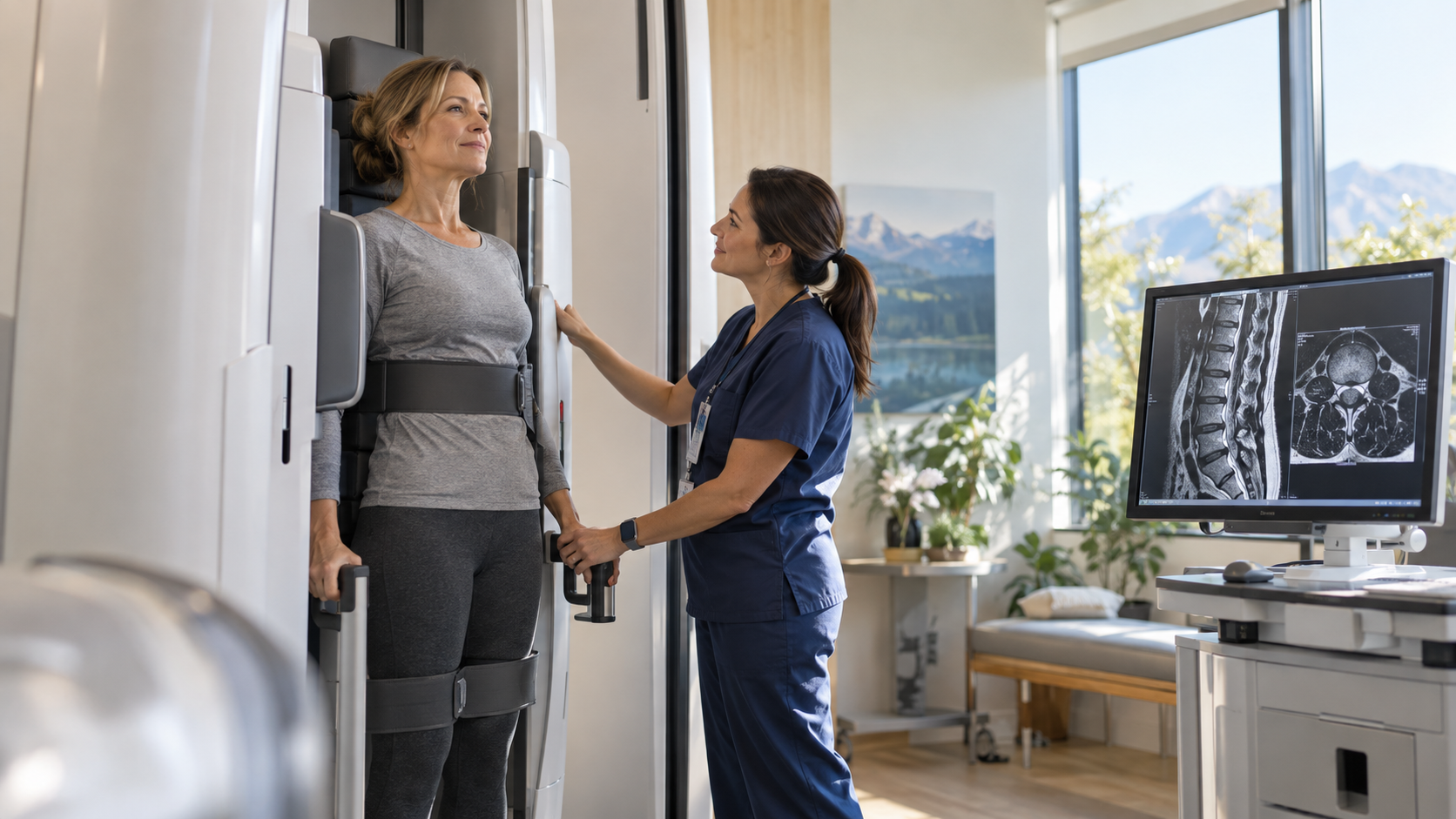
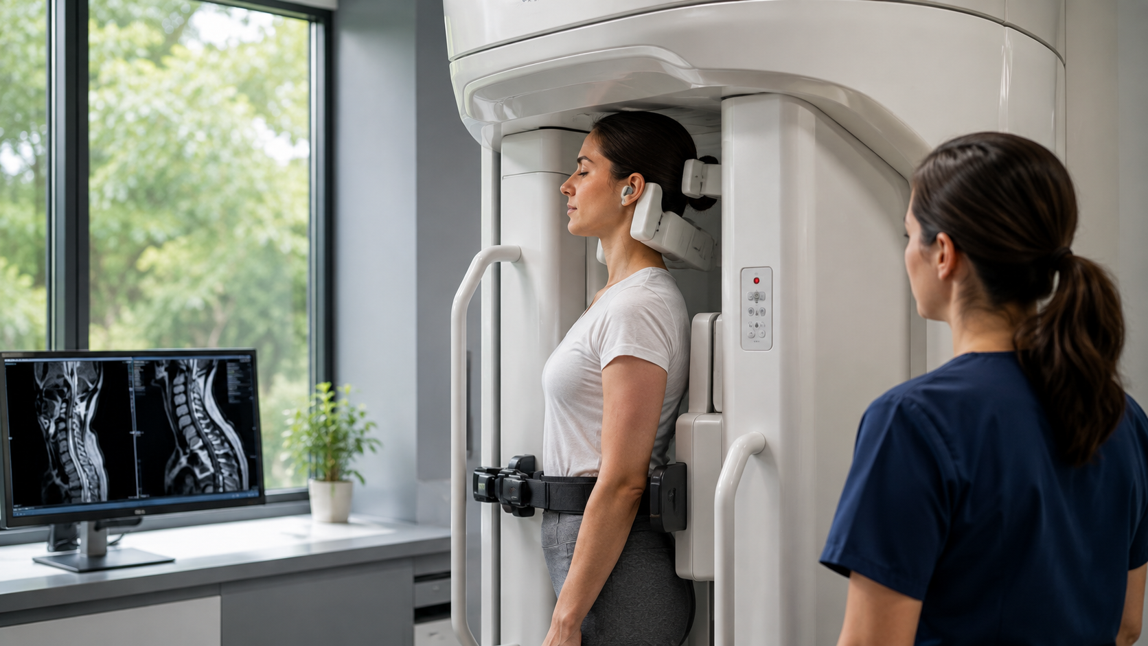
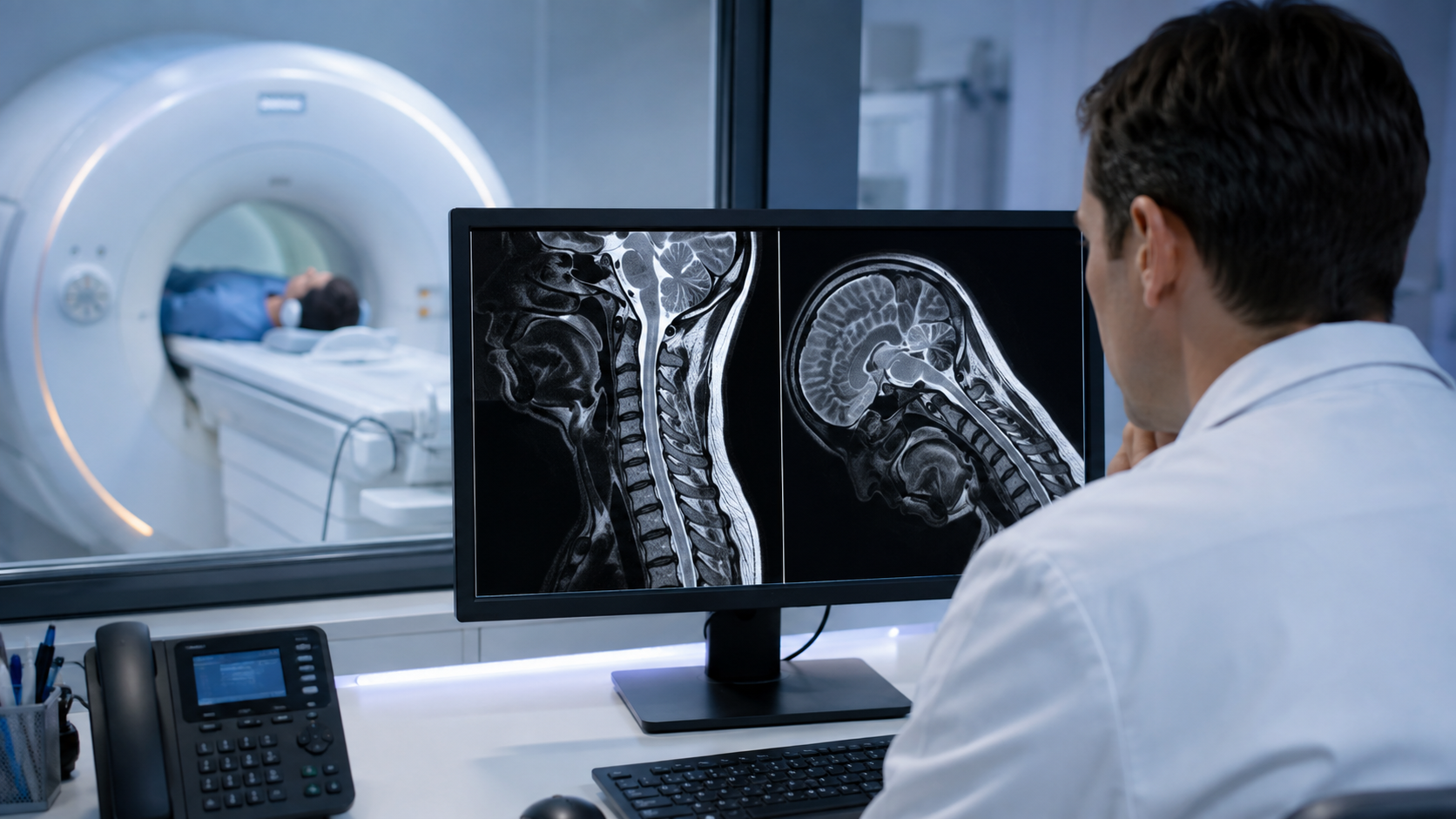
A standard back MRI is usually performed with the patient lying flat. That remains an important and detailed examination, but it does not reproduce every load or posture that causes symptoms. An upright scan gives the radiologist another way to examine the discs while gravity and body position are influencing the spine.
Quick Answer: Yes, an upright back MRI can show some disc bulges more clearly when they become larger or press on nearby structures under body weight. It is especially useful when symptoms worsen while sitting, standing, or changing position. It does not make every disc bulge more visible, so the findings still need to be matched with symptoms, an examination, and the reason the scan was ordered.

Why Can Body Position Change the Appearance of a Disc Bulge?
Spinal discs sit between the vertebrae and help distribute pressure. Their shape is not completely fixed. When a person moves from lying down to sitting or standing, the load across the lumbar spine changes, disc height can shift, and the contour of a bulge can look different on the images.
This is the main value of weight bearing MRI for spine diagnosis. It shows the spine under a condition that is closer to daily life. If a disc moves farther toward the spinal canal or a nerve opening under load, the upright images can document that change.
The difference may be small, moderate, or absent. Upright MRI should therefore be understood as an additional view of spinal mechanics rather than a promise that every abnormality will look worse. A disc that is clearly herniated in all positions may already be well shown on a conventional scan.
What Does a Disc Bulge Look Like on an Upright MRI?
On MRI, a disc bulge appears when disc tissue extends beyond the usual edge of the vertebral bodies. The radiologist looks at its location, width, direction, and relationship to the spinal canal, nerve roots, and neural foramina, which are the openings where spinal nerves leave the spine.
A complete back and spine MRI scan can show discs, nerves, the spinal cord or cauda equina, joints, ligaments, bone marrow, and surrounding soft tissue. Upright views add information about how some of those structures respond when the patient is seated or standing.
Clarity is not only about whether the bulge looks bigger. A more useful scan may show that the bulge approaches a nerve root in one position, reduces space in the canal, or contributes to narrowing at the side of the spine. Those relationships often matter more than the bulge measurement alone.
When Is an Upright MRI Most Likely to Add Useful Detail?
Upright imaging is most relevant when symptoms have a strong positional pattern. If pain or numbness appears while standing but eases after lying down, imaging only in the relaxed position may not capture the same mechanical conditions.
- Symptoms become worse while sitting, standing, walking, bending, or leaning backward.
- A previous lying down MRI did not explain persistent back or leg symptoms.
- The clinician suspects a disc problem, nerve compression, instability, or narrowing that changes with posture.
- The patient cannot comfortably remain flat for a conventional examination.
People with long lasting symptoms may benefit when the imaging position matches the activity that brings on their pain. The discussion of upright MRI for chronic back pain is particularly relevant when earlier testing and the daily symptom pattern do not agree.
Can Flexion or Extension Make a Bulge Easier to See?
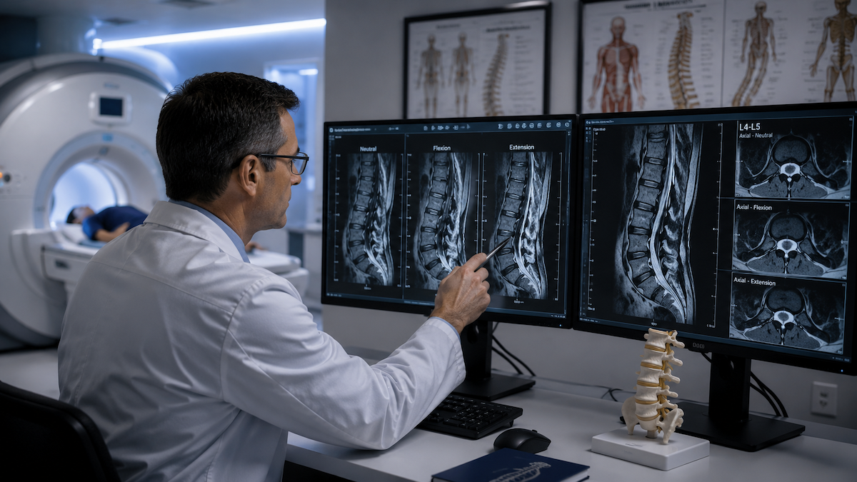
Sometimes. Flexion means bending forward, while extension means leaning backward. Each position changes disc pressure, spinal alignment, and the amount of space around the nerves. A bulge may become more noticeable in one position and less noticeable in another.
A lumbar spine MRI in flexion can help when bending forward is closely connected to symptoms or when movement between vertebrae is a concern. Extension views may be more informative when standing tall or leaning backward triggers pain or leg symptoms.
Comparing neutral and flexion MRI findings can show whether a finding stays stable or changes with posture. This comparison is often more meaningful than treating one position as the single true picture of the spine.
Does a Larger Bulge on Upright MRI Explain the Pain?
Not by itself. Disc bulges are common, and some people have them without pain. A useful interpretation asks whether the level and side of the bulge match the person's symptoms, whether a nearby nerve is affected, and whether other findings could explain the problem better.
For example, leg pain may relate to narrowing where a nerve exits the spine rather than to the overall size of a central bulge. An upright scan may help assess foraminal narrowing under load when the symptoms follow a specific nerve pathway.
The radiology report is one part of the diagnosis. A clinician also considers the history, physical findings, strength, sensation, reflexes, and any warning signs. Imaging is most valuable when all of that information points toward the same explanation.
How Does Upright MRI Compare With a Standard Supine MRI?
A standard supine MRI and an upright MRI answer overlapping but slightly different questions. Supine imaging shows detailed anatomy while the patient is supported and relatively unloaded. Upright imaging shows how that anatomy may change when gravity and posture are added.
- Supine MRI is widely available and remains appropriate for many suspected disc, nerve, infection, tumor, and injury concerns.
- Upright MRI can add functional information when symptoms depend on sitting, standing, flexion, or extension.
- Image quality, field strength, the body area, the protocol, and the patient's ability to remain still all affect the final study.
- The best choice depends on the clinical question, not on a general rule that one scanner is always better.
A normal or mild supine result does not automatically mean another scan is needed. However, flexion MRI after a normal supine scan may be considered when significant, repeatable symptoms remain strongly linked to position and the referring clinician needs more information.
Can Upright MRI Help With Sciatica or Nerve Symptoms?
It can help when sciatica changes with posture. Sciatica describes pain, tingling, numbness, or weakness that travels along the path of a nerve into the buttock or leg. The cause may be a disc herniation, foraminal narrowing, spinal stenosis, or another source of nerve irritation.
A seated MRI for sciatica evaluation can be useful when sitting reliably brings on symptoms. The images may show whether disc shape or the space around a nerve changes in that position.
New loss of bladder or bowel control, numbness around the groin or saddle area, or rapidly worsening leg weakness needs urgent medical assessment. Those symptoms should not wait for a routine imaging appointment.
What Should You Expect From the Results?
The radiologist may compare neutral, seated, standing, flexion, or extension images, depending on the order and the scanner protocol. The report can describe whether a bulge changes in size or shape, whether the canal or foramina narrow, and whether vertebral alignment changes between positions.
The result may confirm a suspected load dependent problem, show that the disc looks similar in every position, or identify a different cause of symptoms. Each outcome is useful because it helps the treating clinician decide what should happen next.
Bring previous scans and reports if the imaging center requests them. Direct comparison can help the radiologist distinguish a new change from a stable finding and can reduce uncertainty when different positions or techniques were used.
Frequently Asked Questions
Can an upright MRI show a disc bulge that a regular MRI missed?
Yes, it can show a position dependent bulge or related narrowing that was less visible while lying down. This is most likely when symptoms appear under load or in a specific posture. It will not uncover a hidden disc problem in every case.
Is an upright MRI better for lower back pain?
It can be more informative when lower back pain changes with sitting, standing, bending, or walking. A standard MRI may still be the right first examination for many patients, depending on the symptoms and the referring clinician's question.
Can a disc bulge look bigger when standing?
Yes. Loading the spine can change disc height and contour, so some bulges look more prominent when seated or standing. Other bulges remain about the same size, which is why comparison between positions matters.
Does an upright MRI hurt?
MRI itself should not hurt, but holding a position may be uncomfortable if that posture triggers symptoms. Tell the technologist what you are feeling so the position and sequence can be managed safely.
Do I need a referral for an upright back MRI?
A physician's order is generally required, and insurance requirements can vary. The order should describe the body area and, when relevant, the positions connected to the symptoms.
Conclusion
An upright back MRI can show certain disc bulges and nerve related changes more clearly when body weight or posture makes them more pronounced. Its greatest value is in answering a specific positional question, while the final diagnosis still depends on the scan, symptoms, and clinical examination together.
Upright MRI of Deerfield provides positional back and spine imaging for patients whose symptoms may change while sitting, standing, or moving. You can visit the practice website or contact the team to discuss the prescription and scheduling process.
SHARE THIS POST:
Leave a Comment:

The World's Most Patient-Friendly MRI. A comfortable, stress-free, and completely reliable MRI scan. We offer patients an open, upright, standup MRI experience that helps those who are claustrophobic and stress being in a confined area. Upright MRI of Deerfield is recognized as the world leader in open MRI innovation,
Our Recent Post

READ PATIENT TESTIMONIALS
Upright MRI of Deerfield.
Susan D.,
Highland Park, 39
I am going to tell everyone about your office! This was a great experience after I panicked in other MRI machines and had to leave. Thank you so much.

Judith B.,
Milwaukee, 61
I suffer from vertigo and other MRIs do not work. This was wonderful…absolutely NO discomfort at all. The MRI was so fast…I wanted to stay and watch the movie! Mumtaz was great. His humor really put me at ease. I’ve already recommended Upright MRI to friends.
Delores P.,
Glencoe, 55
Everything is so nice and professional with your place. I have been there a couple of times. My husband and I would not go anywhere else.