
Do Low Back Pain MRI Results Change When Upright?
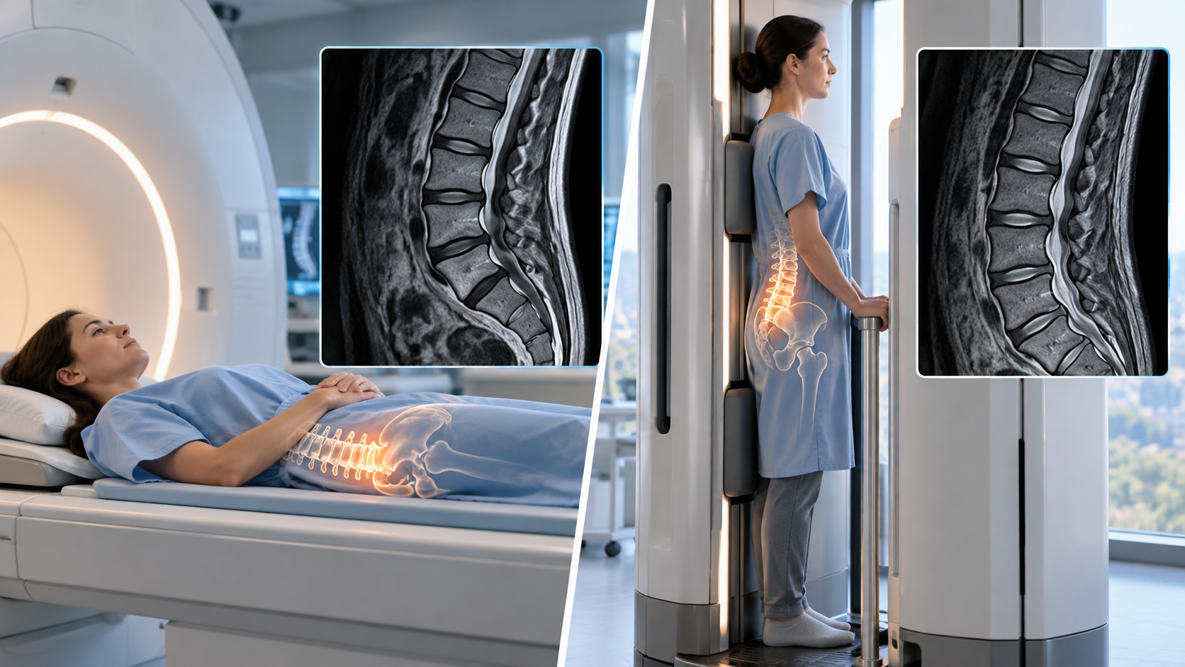
Low back pain is one of the most common reasons adults see a doctor, and MRI is often the test used to look at the discs, nerves, and joints behind it. Most of those scans happen in one position: lying flat on your back with the spine relaxed and unloaded. That is a reasonable way to take a picture, but it is rarely the position in which back pain actually shows up.
Many people notice their pain changes with posture. It builds after standing in line, flares while sitting in the car, or eases the moment they lie down. That pattern raises a fair question for anyone holding a normal or mild MRI report: would the images look different if the scan were taken while standing or sitting, with the spine carrying its usual weight?
Quick Answer: Yes, some low back MRI findings can change when you are upright. Body weight compresses the lumbar spine, which can make disc bulges larger, narrow the spinal canal and nerve openings, and reveal vertebral slipping that relaxes when lying down. Not every finding changes, so upright imaging is most useful when symptoms clearly depend on position.
Why Does Position Matter for a Lumbar MRI?
The lumbar spine is built to carry load. When you stand, the weight of your upper body presses down through the discs and facet joints, and the natural inward curve of the lower back deepens slightly. Lying flat removes most of that load, which gives the discs a chance to settle and the nerves more room.
A conventional MRI captures the spine in that unloaded state. For many conditions this does not matter, because a tumor, infection, or large herniation looks much the same in any position. For problems that are mechanical, though, the unloaded picture can understate what happens when you are on your feet.
That is the reasoning behind weight-bearing spine imaging, which looks at the spine under the same forces that trigger the pain rather than in the position where symptoms often fade.
Which Low Back Findings Can Change When You Stand or Sit?
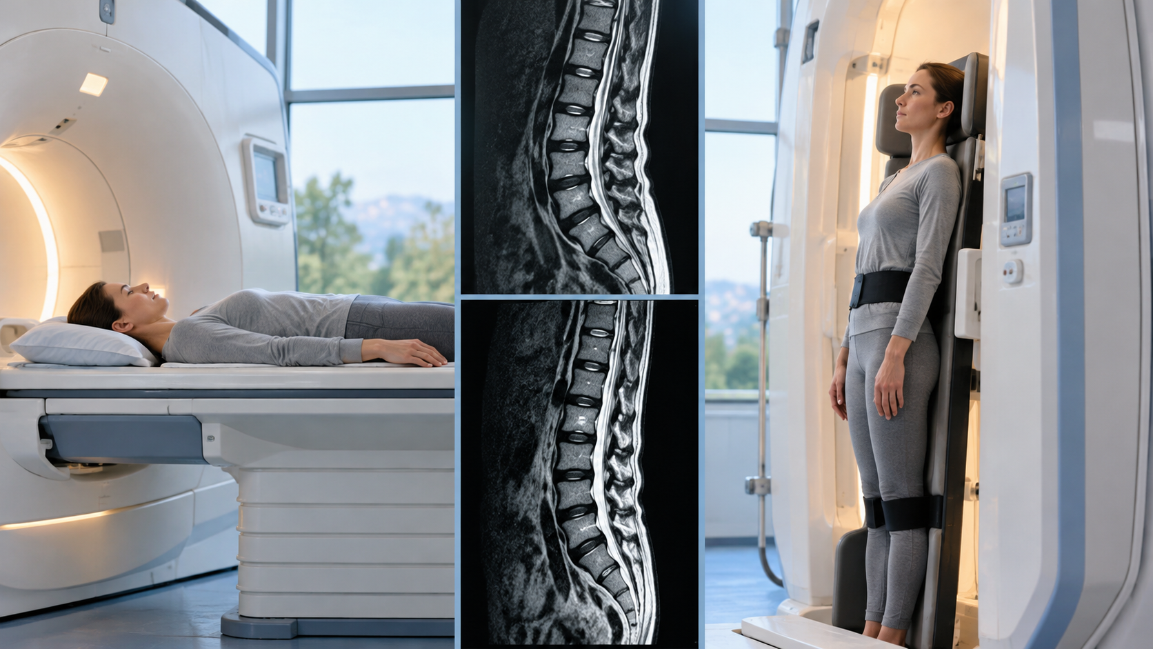
Research on positional and axially loaded MRI has consistently shown that certain measurements shift with posture. The changes are usually measured in millimeters, but in a tight spinal canal a few millimeters can be the difference between a nerve with room to move and a nerve that is pinched.
- Disc bulges and protrusions: under load, a disc bulge can look larger as it pushes back toward the spinal canal.
- Spinal canal size: the canal tends to narrow when standing and when the back arches, which matters for lumbar spinal stenosis.
- Nerve openings: the side openings where nerve roots exit the spine can tighten under load and in extension.
- Vertebral slipping: a spondylolisthesis that partly settles when lying down may show more movement when standing or bending.
- Ligament buckling: the ligament lining the back of the canal can fold inward as the spine shortens under load, adding to narrowing.
What tends not to change is just as important. Disc dehydration, fractures, bone marrow changes, and most structural abnormalities look similar regardless of posture. Upright imaging adds information about how the spine behaves, not a completely different anatomy.
Can a Normal Lying-Down MRI Miss the Cause of Back Pain?
It can, particularly when the pain is dynamic. Studies comparing lying-down and upright scans of the same patients have found that some cases of stenosis, nerve opening narrowing, and instability were only visible, or were clearly worse, when the patient was upright. Those are the findings most likely to explain pain that comes and goes with posture.
The opposite problem also exists. Large reviews of people without back pain show that disc degeneration and bulges are common findings, and they become more common with age. A flat MRI full of abnormal findings does not always point to the true pain source either, which is why matching images to symptoms matters more than the number of findings on a report.
Positional imaging helps close that gap. When a narrowing appears only in the posture that reproduces your pain, it becomes a stronger candidate for the cause than a finding seen at rest that never changes.
Who Is Most Likely to Benefit From an Upright Low Back MRI?
The strongest candidates are people whose symptoms follow a clear positional pattern and whose current imaging does not fully explain them. An upright scan is a targeted tool, so the pattern of your pain is the best guide to whether it will add value.
Leg pain or heaviness that builds while standing or walking and eases when you sit or lean forward is a classic sign of lumbar stenosis. That pattern often responds well to standing images, because the canal is narrowest in exactly that position.
Pain that worsens with prolonged sitting is another common pattern, especially with disc problems and nerve irritation. For these patients, sitting can aggravate sciatica in a way a lying scan never reproduces, and a seated scan captures that loaded posture directly.
People with known or suspected spondylolisthesis, ongoing symptoms after spine surgery, or degenerative disc changes at several levels may also benefit, because each of these involves movement or load that a static, unloaded view can understate.
When Is a Standard Lying-Down MRI Still the Right Choice?
For many back pain cases, a conventional MRI answers the clinical question well. Suspected infection, tumors, fractures, and large disc herniations with clear nerve compression are usually visible without loading the spine, and treatment decisions rarely depend on posture.
Imaging is also not always needed early on. Most new episodes of nonspecific low back pain improve within weeks, and clinical guidelines generally advise against routine imaging in that window unless warning signs appear. Loss of bladder or bowel control, numbness in the groin area, progressive leg weakness, fever, or a history of cancer call for prompt medical care rather than a scheduled scan.
What Happens During an Upright MRI of the Lower Back?
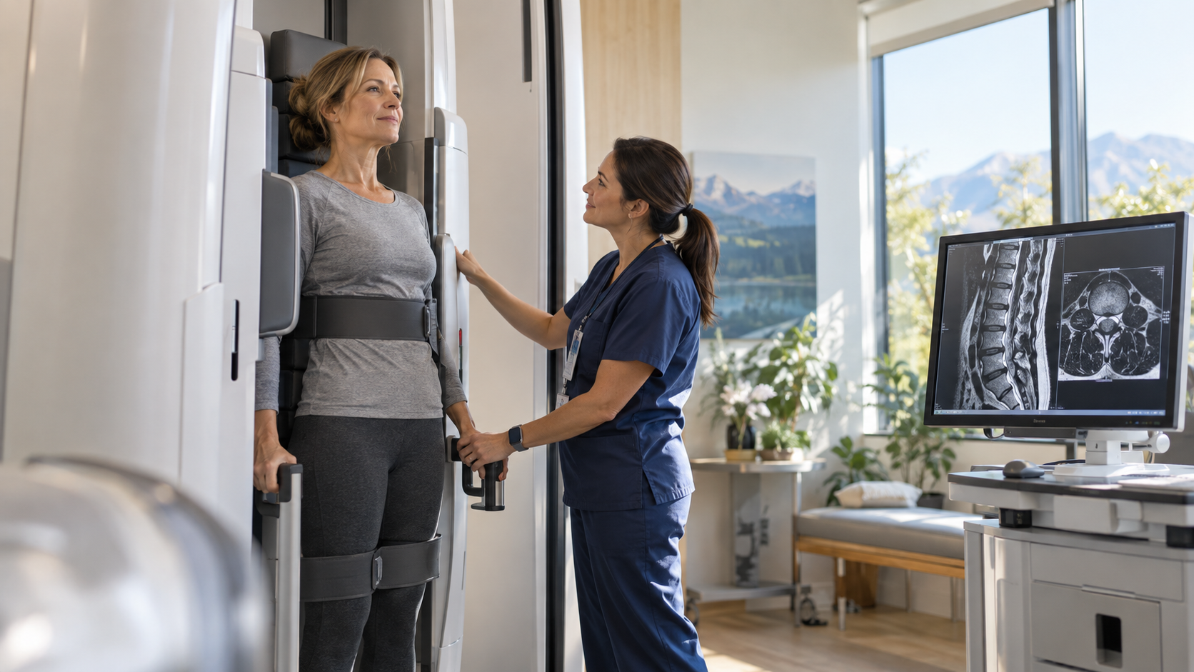
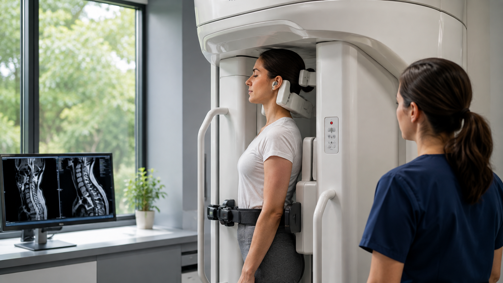
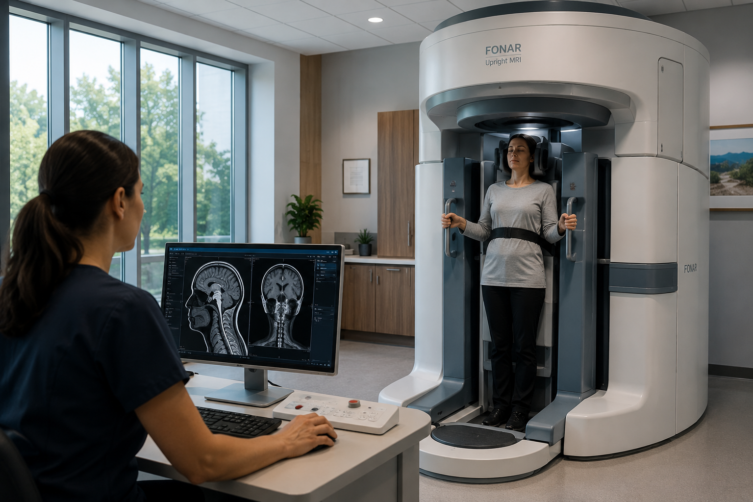
An upright scanner has an open front, so you walk in rather than slide into a tube. Depending on what your doctor orders, the technologist positions you standing, sitting, or bending, and the scanner images the lumbar spine in that posture. You can also be scanned lying down for direct comparison.
Because the setting is open and far quieter than a closed scanner, many people who struggle with traditional MRI find it easier to tolerate. Holding still is still essential, and the technologist uses supports so you can stay steady in each position.
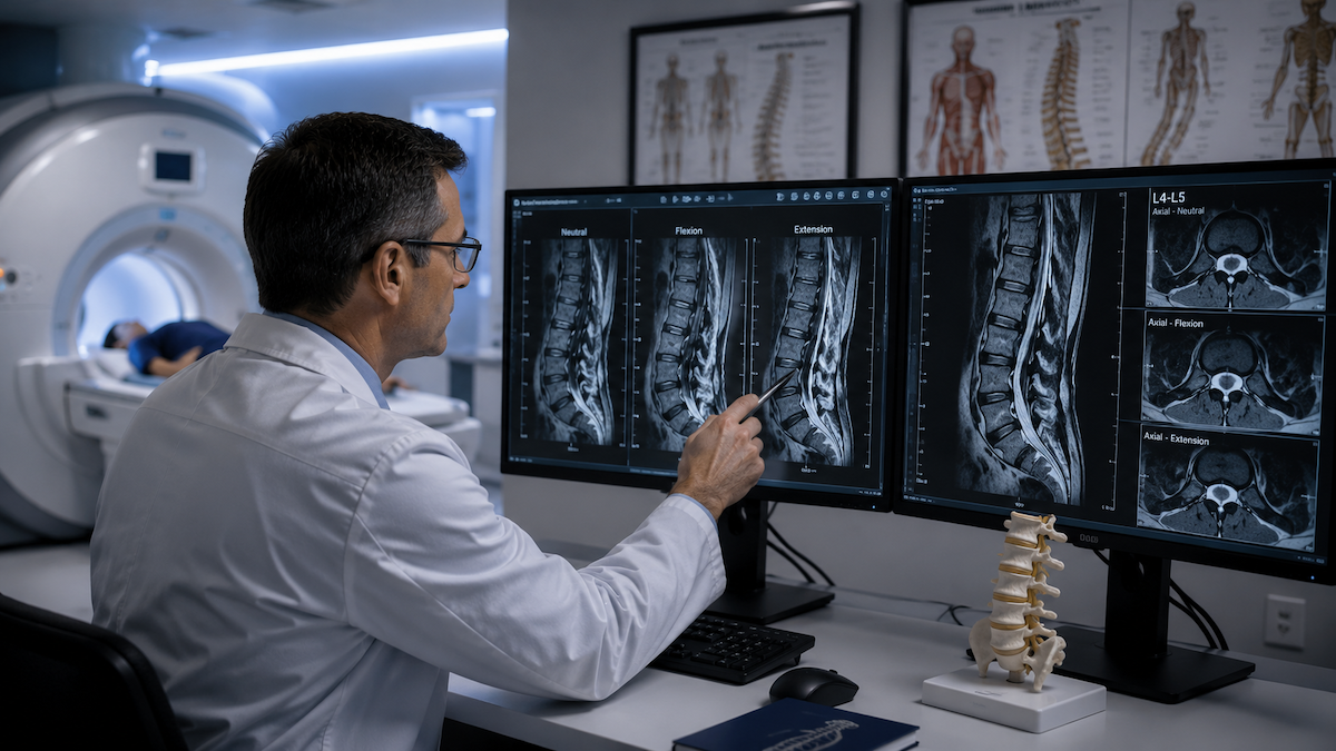
When flexion views are ordered, the images show how vertebrae and discs respond to bending forward in the scanner, which can uncover instability that neutral images miss.
How Should You Talk to Your Doctor About Upright Imaging?
The most useful thing you can bring is a clear description of when your pain appears. Note whether it starts after standing, walking, sitting, or bending, how long it takes to build, and which position relieves it. That detail helps your physician decide whether positional images would change the plan.
If you already had a lying-down MRI, bring the report and images. Your doctor can compare them with your symptoms and decide whether an upright back and spine MRI is likely to answer an open question. A prescription is required, and it can specify which positions to include.
Frequently Asked Questions
Is an upright MRI more accurate than a regular MRI for back pain?
An upright MRI is not more accurate for every condition, but it can be more informative when pain depends on posture. It shows the spine under body weight, which can reveal narrowing, disc bulging, or slipping that lessens when lying down. For infections, tumors, or fractures, a standard MRI is usually sufficient.
Can a herniated disc look different when standing?
Yes, a disc bulge or herniation can push farther toward the spinal canal under load. The change is often a few millimeters, but that can matter when the canal or nerve openings are already narrow. Some herniations look about the same in every position.
Why does my MRI look normal when I am still in pain?
A lying-down MRI shows the spine without the weight and posture that trigger many mechanical pain patterns. If your pain mainly appears when standing, sitting, or bending, the cause may be less visible at rest. Muscles, joints, and nerves outside the spine can also cause pain that a lumbar MRI does not show.
Do I need a referral for an upright lumbar MRI?
Yes, an MRI requires a prescription from a physician. Your doctor can note which positions to scan, such as standing, sitting, or flexion. Bringing previous imaging helps the radiologist compare results.
Does insurance cover upright MRI for low back pain?
Coverage depends on your plan and on how the exam is ordered. Many plans cover lumbar MRI when it is medically necessary, while some limit coverage for added positional views. Confirm with your insurer and the imaging center before your appointment.
Conclusion
Low back pain MRI results can change when you are upright, mostly for mechanical problems like stenosis, nerve opening narrowing, disc bulging, and instability. If your pain clearly follows your posture and a lying-down scan has not explained it, imaging the spine under load may fill in the missing piece.
Upright MRI of Deerfield scans patients standing, sitting, bending, and lying down in an open, quieter setting. If positional imaging sounds right for your symptoms, talk with your doctor and schedule your upright MRI when you are ready.
SHARE THIS POST:
Leave a Comment:

The World's Most Patient-Friendly MRI. A comfortable, stress-free, and completely reliable MRI scan. We offer patients an open, upright, standup MRI experience that helps those who are claustrophobic and stress being in a confined area. Upright MRI of Deerfield is recognized as the world leader in open MRI innovation,
Our Recent Post


READ PATIENT TESTIMONIALS
Upright MRI of Deerfield.
Susan D.,
Highland Park, 39
I am going to tell everyone about your office! This was a great experience after I panicked in other MRI machines and had to leave. Thank you so much.

Judith B.,
Milwaukee, 61
I suffer from vertigo and other MRIs do not work. This was wonderful…absolutely NO discomfort at all. The MRI was so fast…I wanted to stay and watch the movie! Mumtaz was great. His humor really put me at ease. I’ve already recommended Upright MRI to friends.
Delores P.,
Glencoe, 55
Everything is so nice and professional with your place. I have been there a couple of times. My husband and I would not go anywhere else.