
Why Use a Seated MRI for Sciatica Evaluation?
Sciatica is one of the most disruptive pain conditions a person can experience. The shooting, burning, or electric sensation that travels from the lower back down through the buttock and into the leg can make sitting, standing, walking, and even lying down deeply uncomfortable. For the millions of people living with it, getting an accurate picture of what is actually causing the problem is the essential first step toward real relief.
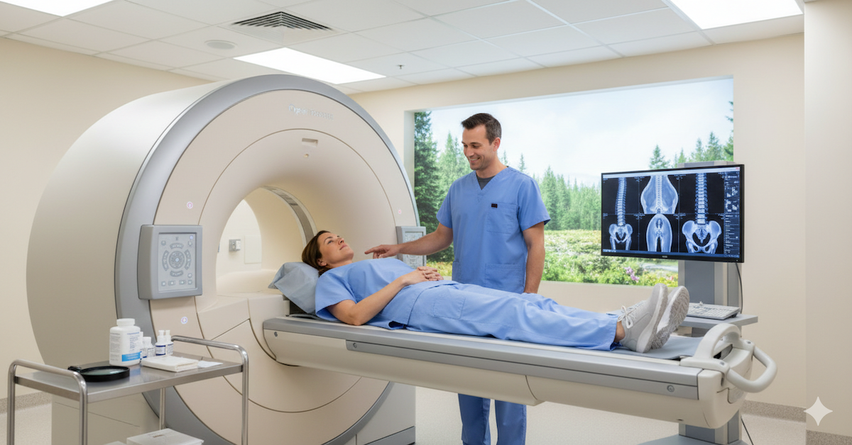
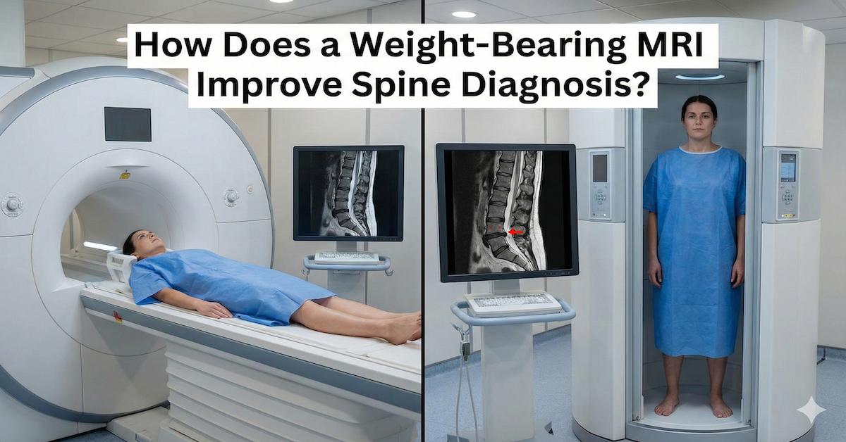
Most people with sciatica are referred for a traditional MRI as part of their diagnostic workup. That scan is usually performed lying flat on a table, in a position where the spine is fully supported and the muscles around it are relaxed. The problem is that sciatica almost never feels worst when you are lying down. It flares when you sit, when you stand, when you walk. The question worth asking is whether a scan taken in a completely different position from the one that triggers symptoms will actually capture what is causing them.
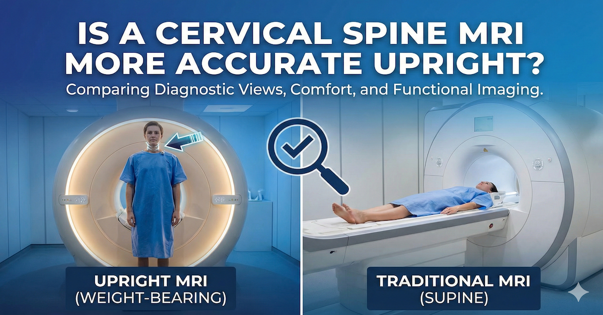
Quick Answer: A seated MRI is valuable for sciatica evaluation because it images the spine under the weight-bearing conditions that actually provoke symptoms. When a person sits or stands, gravitational load compresses the lumbar discs and narrows the spaces where spinal nerves travel. A scan taken in this position is far more likely to reveal the nerve compression or disc bulging driving the sciatica than one taken lying flat, where those same structures may decompress and appear normal.

The Position Problem With Conventional Sciatica Imaging
Sciatica is almost always a positional condition. The nerve irritation that causes it typically results from a structure pressing on or narrowing the path of the sciatic nerve, and that compression is usually made worse by loading the spine. Sitting places significant pressure on the lumbar discs. Standing adds compressive load. Walking involves dynamic forces across the entire lower back and pelvis. These are the circumstances under which the nerve gets squeezed.
When you lie flat for a conventional MRI, the weight is removed from the spine entirely. Muscles relax, discs partially decompress, and the structures that were encroaching on nerve tissue may shift enough to look substantially different from how they appear when you are upright. This limitation of lying-down imaging is well documented and directly affects diagnostic accuracy for positional conditions like sciatica.
The clinical consequence is that patients sometimes receive a report describing mild or even normal findings, when in practice they are in significant pain during any upright activity. When what the scan says does not match what the patient feels, the path to effective treatment becomes much harder to navigate.
How Seated Positioning Changes the Diagnostic Picture
A seated MRI places the patient in a position that actually loads the lumbar spine. Gravity acts on the discs and facet joints in a way that reflects everyday function. When this load is applied, several things can become visible that remain hidden in a lying-down scan.
Disc bulges and herniations may enlarge or shift position under load, pressing more significantly into the nerve canal. The foramina, the small openings through which nerve roots exit the spinal column, can narrow noticeably when the spine is compressed. Ligaments at the back of the spinal canal can buckle inward. Subtle instability between vertebral segments can become apparent. All of these findings have direct relevance to sciatica, and all of them are best captured when the spine is bearing weight.
This is why weight-bearing spine imaging provides information that simply cannot be replicated by taking a person out of the symptomatic position and scanning them at rest.
Sciatica and Nerve Compression: Why Position-Sensitive Imaging Matters
The sciatic nerve is formed from several nerve roots that exit the lower lumbar spine, typically between L4 and S1. Any structure that compresses these roots, whether a disc, a bone spur, or a narrowed foramen, can trigger the characteristic radiating pain down the leg. The degree of that compression can change significantly depending on spinal position.
For patients whose symptoms are clearly worse when sitting, a seated MRI is particularly revealing. The seated position loads the lower lumbar segments most intensively, and this is where the vast majority of sciatica-causing pathology originates. Studies comparing upright and supine MRI findings consistently show that upright imaging detects more in a meaningful proportion of patients whose lying-down scan appeared normal or only mildly abnormal.
Who Should Consider a Seated MRI for Sciatica
A seated MRI is particularly useful for certain groups of patients. These include people whose sciatica or leg pain is clearly worse when upright and improves when lying down, people who have had a conventional MRI that did not find adequate explanation for their symptoms, and people whose treatment plan has not been effective despite a seemingly complete workup.
It is also highly relevant for patients with symptoms that come and go with activity. Dynamic instability between spinal segments, which can cause intermittent nerve irritation, is essentially invisible in a supine scan but may become apparent when the spine is imaged under load.
For anyone living with chronic back-related leg pain and seeking a clearer explanation, this overview of upright MRI for back pain outlines the broader case for positional imaging across different types of persistent spinal conditions.
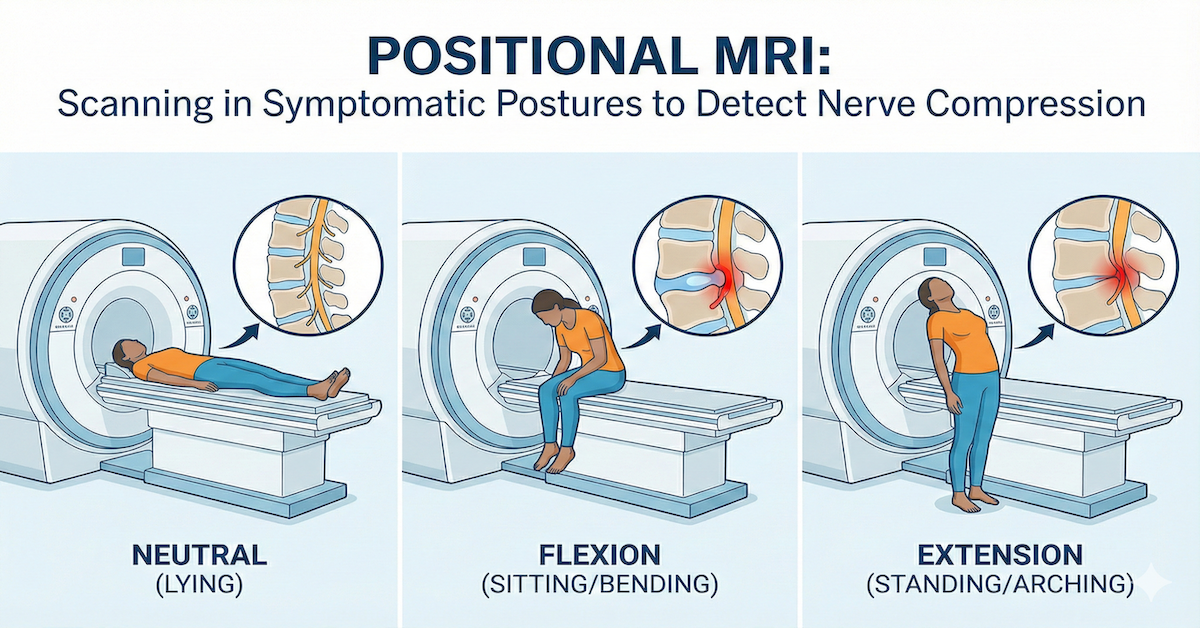
Flexion Imaging and Its Role in Sciatica Diagnosis
One of the unique advantages of an upright MRI is the ability to image the spine in flexion and extension, meaning bent forward and arched backward. These positions can reveal instability and nerve encroachment that is completely absent in a neutral scan.
For sciatica patients who notice their symptoms change significantly depending on posture, lumbar flexion imaging can identify which specific spinal position triggers the nerve compression, giving treating clinicians far more targeted information to work with.
A physiotherapist, pain specialist, or surgeon working from positional imaging data has a clearer understanding of what is actually happening and when. Treatment decisions become more precise and the likelihood of the right intervention improves considerably.
What the Scan Experience Is Like
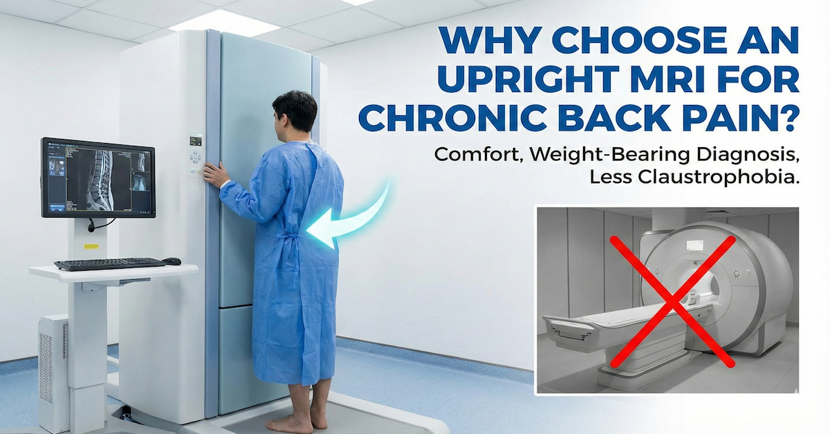
An upright MRI for sciatica evaluation involves sitting in a chair-like position within the scanner. There is no tunnel to enter and no need to lie flat. The open design of the machine means the experience is considerably more comfortable for many patients, particularly those who find conventional scanner environments claustrophobic or who have difficulty lying flat for extended periods due to their pain.
The scan itself takes a similar length of time to a conventional lumbar MRI. The images are then reported by a radiologist who reviews them in the context of the patient's symptomatic positions. For many patients this is the first time a scan truly reflects what their body is doing when it hurts.
More detail on what is covered and how the process works is available on the back and spine scan page at Upright MRI of Deerfield.
Frequently Asked Questions
Is a seated MRI always better than a lying-down MRI for sciatica?
Not in every case, but for patients whose sciatica is position-dependent, a seated or upright MRI is significantly more likely to identify the source of the problem. If a lying-down scan has already returned clear findings but symptoms persist, a seated scan is often the logical next step.
Will a seated MRI show the same things as a traditional MRI?
A seated MRI can show all the same structures as a traditional scan but with the added dimension of gravitational load. In many cases it reveals additional findings such as increased disc bulging, greater foraminal narrowing, or vertebral instability that are not visible when the spine is unloaded.
Can I request a seated MRI if my doctor ordered a standard one?
Yes. Patients can self-refer for an upright MRI independently of a physician referral at many facilities. The resulting images and report can be shared with your treating physician to inform the next steps in your care.
How is sciatica different from general back pain, and does it change what scan I need?
Sciatica specifically involves radiating pain along the path of the sciatic nerve, typically down one leg. Because it is caused by nerve compression rather than simple muscle strain, imaging that reveals the degree and location of that compression under load is more diagnostically useful than imaging taken at rest.
Does insurance cover a seated or upright MRI for sciatica?
Coverage varies by plan and provider. Some insurers cover upright MRI under standard MRI benefits while others do not. Self-pay options are available at many upright MRI facilities, and the cost is often comparable to or lower than out-of-pocket costs at hospital-based imaging centres.
The Bottom Line
Sciatica is a condition that lives in upright positions, and imaging it lying down does not always capture the full picture. A seated MRI evaluates the lumbar spine under the weight-bearing conditions that trigger symptoms, giving physicians and patients a more accurate and actionable set of findings.
Upright MRI of Deerfield specialises in exactly this type of imaging. If your sciatica has not been adequately explained by previous imaging, reaching out to the team is a straightforward next step toward understanding what is really going on.
SHARE THIS POST:
Leave a Comment:

The World's Most Patient-Friendly MRI. A comfortable, stress-free, and completely reliable MRI scan. We offer patients an open, upright, standup MRI experience that helps those who are claustrophobic and stress being in a confined area. Upright MRI of Deerfield is recognized as the world leader in open MRI innovation,
Our Recent Post


READ PATIENT TESTIMONIALS
Upright MRI of Deerfield.
Susan D.,
Highland Park, 39
I am going to tell everyone about your office! This was a great experience after I panicked in other MRI machines and had to leave. Thank you so much.

Judith B.,
Milwaukee, 61
I suffer from vertigo and other MRIs do not work. This was wonderful…absolutely NO discomfort at all. The MRI was so fast…I wanted to stay and watch the movie! Mumtaz was great. His humor really put me at ease. I’ve already recommended Upright MRI to friends.
Delores P.,
Glencoe, 55
Everything is so nice and professional with your place. I have been there a couple of times. My husband and I would not go anywhere else.

