Does an Upright Shoulder MRI Show Tears More Clearly?
Shoulder pain that persists after rest, physical therapy, and conservative treatment eventually leads most patients and clinicians to imaging. An MRI is the preferred tool for evaluating the soft tissues of the shoulder, and in many straightforward cases a conventional supine scan provides exactly what is needed. But the shoulder is not a passive joint. Its function is almost entirely load-bearing and positional, and the structures most commonly involved in shoulder pain, the rotator cuff tendons, the labrum, and the surrounding bursae, behave differently depending on whether the arm is at the side, raised, or bearing weight.
This creates a genuine diagnostic gap. A scan taken with the patient lying flat, arm relaxed at the side, captures the anatomy in a position that most symptomatic shoulder patients rarely occupy in real life. Someone whose pain is worst when reaching overhead, lifting objects, or pressing with the arm outstretched is describing a dynamic, load-dependent problem that may not fully reveal itself on a static resting scan.
Quick Answer: An upright shoulder MRI can show tears and joint pathology more clearly than a supine scan in cases where the abnormality is positional or load-dependent. Imaging the shoulder in the seated or standing position, with the arm in the position that provokes symptoms, places the rotator cuff tendons, labrum, and bursa under real functional load. This can reveal tears, impingement patterns, and instability that are absent or significantly underrepresented on a supine study.
Why the Shoulder Joint Is Load-Sensitive
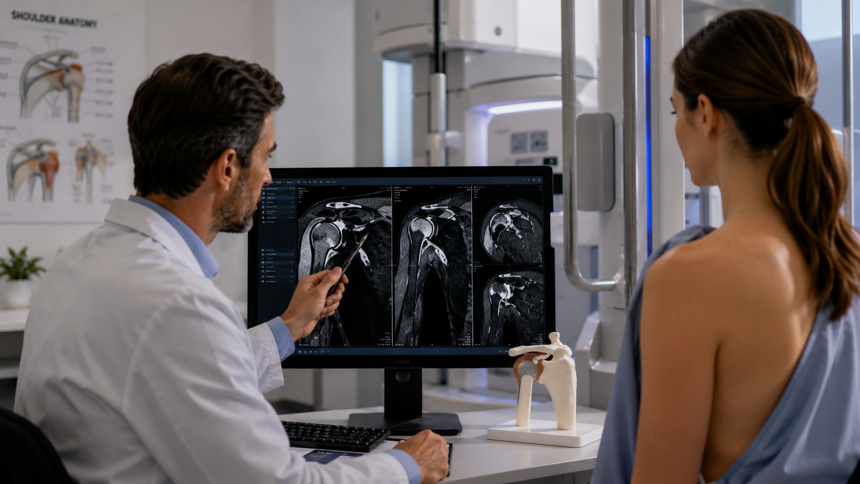
The glenohumeral joint, the ball-and-socket at the core of the shoulder, is uniquely dependent on soft tissue for its stability. Unlike the hip, where the bony socket provides substantial mechanical containment, the shoulder trades bony stability for a massive range of motion. The rotator cuff tendons, the labrum, and the joint capsule together provide the dynamic stabilisation that keeps the joint functional.
What this means for imaging is that the appearance of these structures changes with arm position and loading. A rotator cuff tendon that appears intact at rest may show thinning, fraying, or a partial tear when the arm is elevated or placed under the kind of torque generated by overhead activity. The subacromial space, where impingement commonly occurs, narrows and shifts with arm elevation in ways that a neutral resting position does not capture.
The shoulder is one of the joints where positional MRI reveals hidden issues most consistently, because the dynamic functional demands placed on the shoulder differ so substantially from the resting anatomy a supine scan captures.
What Supine Shoulder MRI Misses
A standard supine shoulder MRI with the arm at the side produces excellent images of many types of pathology. Full-thickness rotator cuff tears with significant retraction, calcific tendinitis, large bony abnormalities, and tumours all present clearly regardless of position. For these diagnoses, supine imaging is entirely adequate.
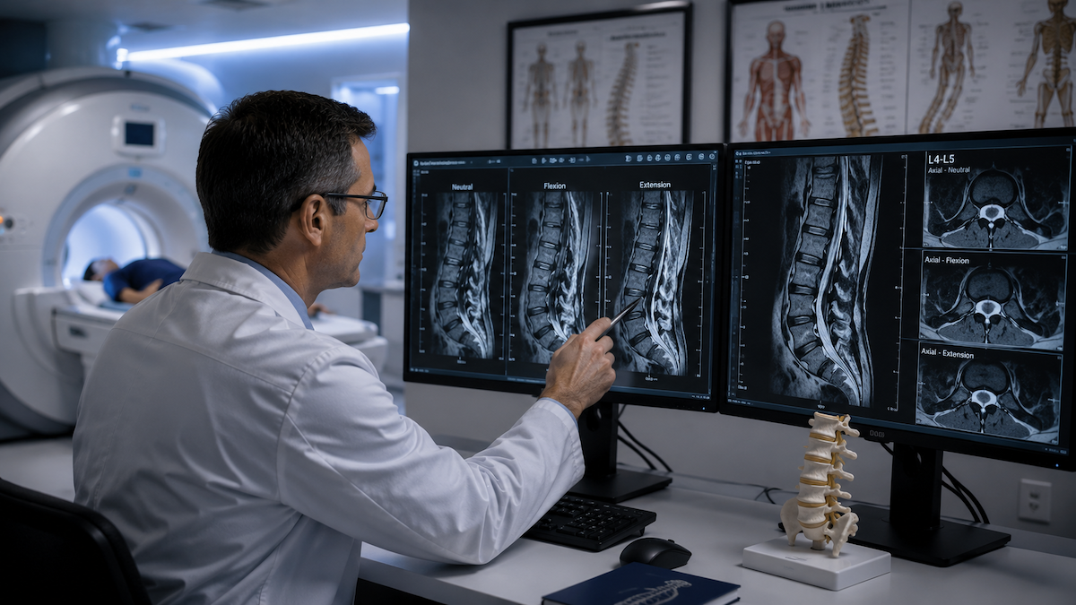
The category where supine imaging falls short involves pathology that is positional or dynamic. Partial-thickness tears that open or become more apparent under loading may be subtle or absent on a resting scan. Superior labral tears in overhead athletes often produce the most significant abnormality when the arm is in the position of maximum stress rather than at rest. Subacromial impingement, by definition a dynamic problem caused by narrowing of the subacromial space during arm elevation, is difficult to image in a position that avoids the elevation producing the impingement.
This is the core limitation that supine MRI is not enough to address in cases where symptoms are clearly linked to specific arm positions or loading activities that the supine position does not reproduce.
Rotator Cuff Tears: How Upright Imaging Adds Information
The four rotator cuff muscles, the supraspinatus, infraspinatus, teres minor, and subscapularis, each have different vulnerability profiles and different positional characteristics. The supraspinatus tendon, which passes through the subacromial space during arm elevation, is the most commonly torn and also the most affected by subacromial narrowing that occurs with arm movement.
In an upright MRI with the arm positioned at shoulder height or above, the supraspinatus is placed under the mechanical tension and positional stress it experiences during the activities that most commonly produce symptoms. A partial tear that appears as mild tendon signal change on a resting scan may show clear fibre disruption, fluid ingress, or width reduction when the tendon is loaded and positioned correctly.
For patients with clinical findings strongly suggestive of a rotator cuff tear but a supine MRI reported as normal or equivocal, upright positional imaging in the symptomatic position represents a logical next diagnostic step before either accepting a false-negative result or proceeding to more invasive assessment.
Labral Tears and Shoulder Instability
The labrum is the fibrocartilaginous ring that deepens the shallow glenoid socket and provides an attachment point for the glenohumeral ligaments. Labral tears are a significant source of shoulder pain and instability, particularly in younger, active individuals and overhead athletes. The specific type and location of the tear has direct implications for the treatment approach and the likelihood of surgical versus conservative management.
Superior labral tears from anterior to posterior, known as SLAP lesions, are notoriously difficult to image reliably on standard supine MRI. The relatively thin tissue involved and the close proximity of normal anatomical variants to pathological findings means that supine imaging has meaningful false-negative and false-positive rates for this particular tear type. Positioning the shoulder in the abducted and externally rotated position during imaging places the superior labrum under the stress that most commonly reveals the tear.
Patients with recurrent shoulder instability and episodes of subluxation or dislocation present a different imaging challenge. The anatomical changes associated with instability, stretching of the capsular ligaments, bone bruising at specific contact points, and labral detachment, may be best appreciated when the joint is in the position of instability risk rather than in a neutral resting state.
Subacromial Impingement: A Fundamentally Positional Diagnosis
Subacromial impingement describes the compression of the supraspinatus tendon and the subacromial bursa between the humeral head and the acromion during arm elevation. By definition, it is a position-dependent phenomenon that requires arm movement to produce the impingement. Imaging in a position that avoids arm elevation does not capture the problem at its most apparent.
An upright MRI that images the shoulder in progressive degrees of arm elevation can visualise the subacromial space narrowing that occurs with the movement, the position of the tendon within the space, and whether significant bursal thickening or fluid accumulation accompanies the impingement. This information helps confirm the mechanism of pain and can influence whether conservative management.
subacromial injection, or surgical decompression is the most appropriate direction.
The same principle of weight-bearing MRI improves diagnosis by capturing joints under real functional conditions applies directly to the shoulder, where the relevant functional conditions involve gravitational load and arm position rather than simply body weight.
Nerve Compression Around the Shoulder
The brachial plexus and peripheral nerves around the shoulder can be compressed by positional changes in the cervical spine and shoulder complex, and these compressions may only manifest when the arm is in specific positions. Nerve compression detection in the shoulder region benefits from positional imaging for the same reason it does in the spine: the anatomy changes with position, and the compression that produces symptoms may not be present in a neutral resting state.
Thoracic outlet syndrome, in which neurovascular structures are compressed between the clavicle and first rib during arm elevation, is one example where positional imaging provides diagnostic information unavailable from a resting study. Imaging with the arm elevated in the provocative position can demonstrate the compression that correlates with the patient's reported symptoms.
Who Should Consider Upright Shoulder Imaging
Upright positional shoulder MRI is not necessary for every shoulder complaint. For straightforward cases with a clear clinical diagnosis supported by a normal or clearly abnormal supine study, additional positional imaging adds little. The patients who benefit most are those whose clinical picture does not match their supine MRI findings, who have position-dependent or activity-dependent pain that the resting scan has not explained, or who are being considered for surgery on the basis of imaging that may not capture the full extent of the pathology.
Overhead athletes with superior labral symptoms, patients with recurrent shoulder instability, workers or athletes whose pain is specifically provoked by loaded arm positions, and patients with an equivocal supine MRI that has not resolved the clinical question are all groups where an upright positional study adds meaningful diagnostic value.
Referring clinicians considering positional shoulder imaging will find physician referral information at Upright MRI of Deerfield useful for understanding the protocols available and how to request studies that target the specific positional scenarios relevant to each patient.
Frequently Asked Questions
Is an upright shoulder MRI the same as an MRI arthrogram?
No. An MRI arthrogram involves injecting contrast fluid into the joint before scanning to distend the capsule and improve labral visibility. An upright positional MRI images the shoulder in different positions without any injection. The two approaches provide different types of information and are sometimes used complementarily.
Can an upright MRI diagnose a full rotator cuff tear?
Yes. Full-thickness rotator cuff tears with retraction are visible on both supine and upright MRI. The upright approach adds most value for partial tears, positional impingement, and labral pathology where the resting position underrepresents the severity of the finding.
Is the upright shoulder MRI more uncomfortable than a standard scan?
Some patients find the upright position more comfortable because they are seated rather than lying inside a narrow tube. The arm positioning required for specific shoulder protocols may involve some discomfort for people with significant shoulder pain, but most patients tolerate the study well. Staff can adjust positioning to balance diagnostic value with patient comfort.
Will my insurance cover an upright shoulder MRI?
Coverage varies by insurer and by the clinical justification provided. Positional MRI is increasingly recognised as clinically appropriate for specific indications. Providing a clear rationale for why conventional imaging was insufficient and what specific clinical question the positional study is intended to answer strengthens the case for coverage.
Can shoulder MRI be performed in the upright position in a standard scanner?
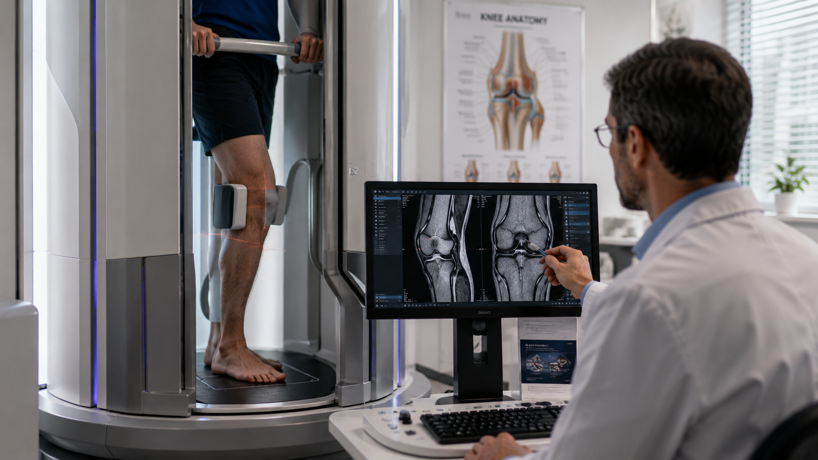
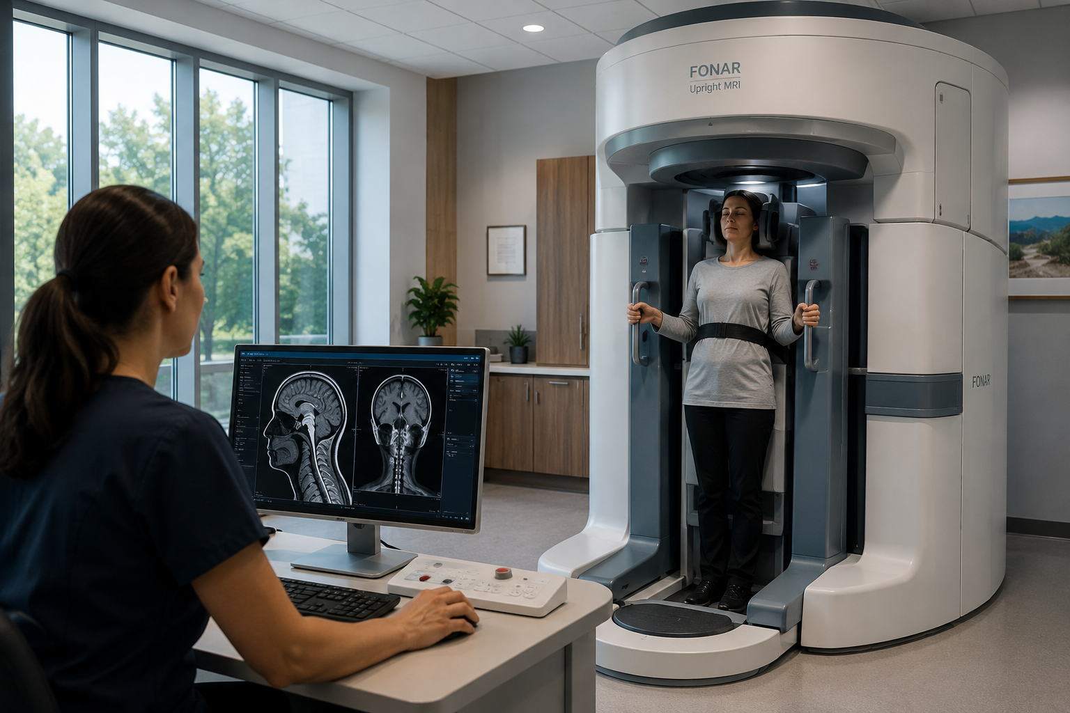
Standard closed-bore MRI scanners require the patient to lie supine, which prevents true upright or seated shoulder imaging. Upright open MRI systems that allow the patient to be scanned in a seated or standing position with the arm in various orientations are specifically designed for this purpose.
The Bottom Line
An upright shoulder MRI provides clinically valuable information that a supine scan cannot when the pathology is positional, load-dependent, or most apparent with the arm in specific orientations. Rotator cuff tears, labral lesions, and subacromial impingement are all conditions where the functional position of the shoulder matters for accurate imaging.
Upright MRI of Deerfield provides positional shoulder imaging for patients and referring clinicians in the Chicago area and Midwest. If a conventional scan has not explained your shoulder symptoms, a consultation is a practical next step.
SHARE THIS POST:
Leave a Comment:

The World's Most Patient-Friendly MRI. A comfortable, stress-free, and completely reliable MRI scan. We offer patients an open, upright, standup MRI experience that helps those who are claustrophobic and stress being in a confined area. Upright MRI of Deerfield is recognized as the world leader in open MRI innovation,
Our Recent Post
READ PATIENT TESTIMONIALS
Upright MRI of Deerfield.
Susan D.,
Highland Park, 39
I am going to tell everyone about your office! This was a great experience after I panicked in other MRI machines and had to leave. Thank you so much.

Judith B.,
Milwaukee, 61
I suffer from vertigo and other MRIs do not work. This was wonderful…absolutely NO discomfort at all. The MRI was so fast…I wanted to stay and watch the movie! Mumtaz was great. His humor really put me at ease. I’ve already recommended Upright MRI to friends.
Delores P.,
Glencoe, 55
Everything is so nice and professional with your place. I have been there a couple of times. My husband and I would not go anywhere else.