
Does an Upright MRI Reveal Foraminal Narrowing Better?
Foraminal narrowing is one of the most frequently cited causes of radiating nerve pain in the neck and back, yet it remains one of the most commonly underdiagnosed spine conditions. Patients often go through multiple rounds of imaging, various treatments, and sometimes even surgery consultations before someone identifies that the small openings through which their spinal nerve roots exit are the source of the problem. Part of the reason this happens is how and where those openings are imaged.
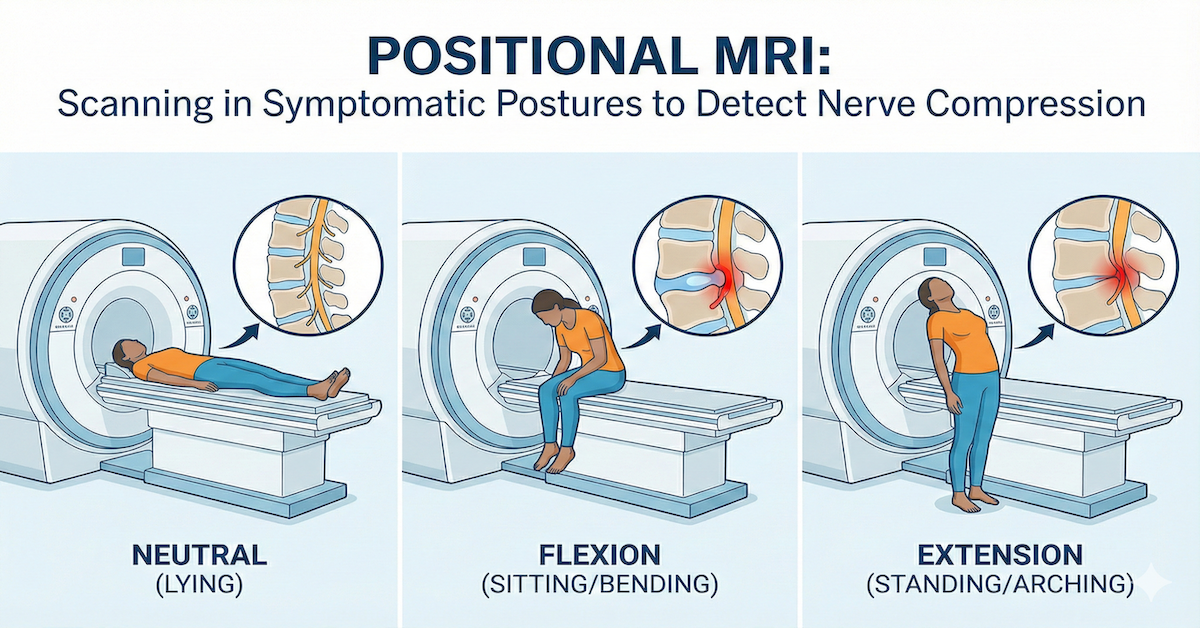
The intervertebral foramina are tiny channels on the sides of the spinal column, one on each side at every vertebral level. Each one provides a pathway for a nerve root to travel from the spinal cord out into the body. When those channels narrow, whether through disc degeneration, bone spur formation, or ligament thickening, the nerve inside can become compressed, producing pain, numbness, tingling, or weakness that radiates along the nerve's path. Understanding the extent of that narrowing depends heavily on how the spine is positioned when the imaging takes place.
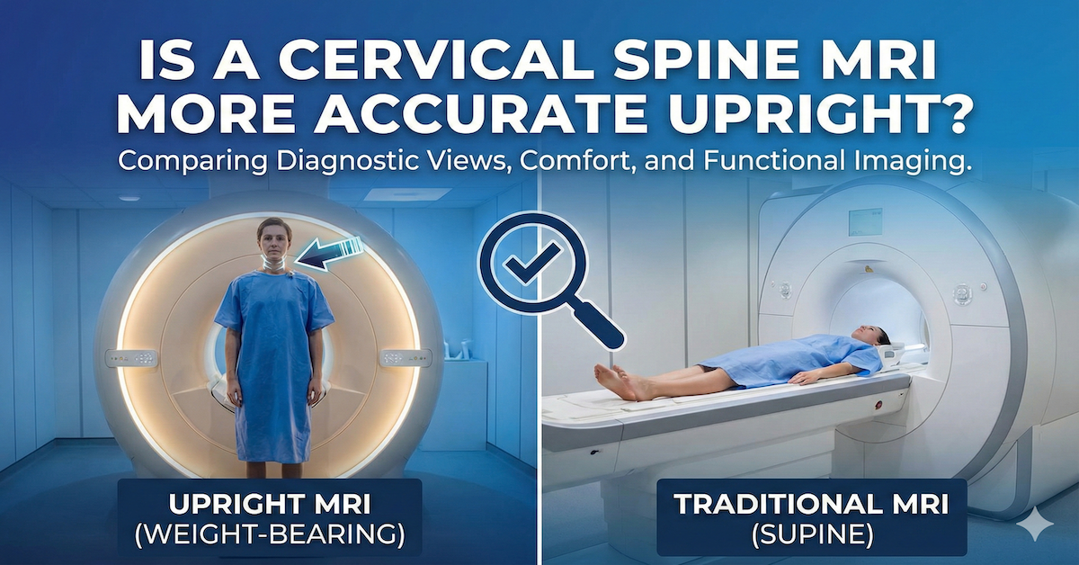
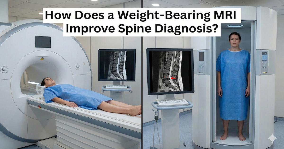
Quick Answer: Yes, an upright MRI typically reveals foraminal narrowing more accurately than a lying-down scan. The foramina change size and shape depending on spinal load and position. When a person is standing or seated, gravitational forces compress the spine and can significantly reduce the size of these nerve openings. Imaging the spine in this loaded state captures the true degree of narrowing that patients experience during daily activities, rather than the partially decompressed state seen when lying flat.

What Happens to the Foramina When You Stand or Sit
The intervertebral foramen is not a fixed structure. Its dimensions change with every movement of the spine and with the presence or absence of gravitational load. When the spine is loaded, as it is when sitting, standing, or walking, the discs between the vertebrae compress slightly, the vertebral segments move closer together, and the foraminal height can decrease measurably. If there is already some degree of degenerative change in that area, this load-related narrowing can push the foramen from a borderline measurement to one that is actively compressing the nerve inside it.
When a person lies down, the opposite happens. The load is removed, the discs partially rehydrate and expand, the vertebral segments separate slightly, and the foramina open up. A structure that was critically narrow in a standing or sitting position may appear within normal limits in a supine scan. This is not an error in the imaging; it is a genuine physiological change that means a lying-down scan may significantly underestimate the degree of nerve compression a patient is experiencing during everyday activities.
This positional dependency is at the core of why upright MRI has a diagnostic advantage for load-sensitive spinal conditions like foraminal stenosis.
The Weight-Bearing Effect on Foraminal Dimensions
Research into positional changes in the lumbar and cervical spine has consistently demonstrated that foraminal cross-sectional area decreases in extension and when axial load is applied. In the lumbar spine, studies using upright MRI have found that foraminal area can be significantly reduced in standing and seated positions compared to supine, with some measurements showing reductions substantial enough to change the clinical significance of the finding entirely.
This means a foramen that appears adequately sized in a lying-down MRI can appear critically narrowed in a weight-bearing scan of the same patient. The clinical value of detecting this difference is substantial, and the mechanics behind it explain why load changes so much of what the spine reveals on imaging.
In the cervical spine, the foramina are smaller to begin with and are affected by both neck extension and axial loading. Patients whose neck and arm pain is worst when looking upward or carrying weight are experiencing exactly this type of positional foraminal compression. Imaging them in that provocative position rather than lying still in a neutral neck position is a fundamentally more informative diagnostic approach.
Cervical Foraminal Narrowing and Upright Imaging
Cervical foraminal narrowing is a frequent cause of arm pain, numbness, and hand weakness, and it is a condition where the benefit of upright imaging is particularly well supported. Many patients with this type of nerve root irritation find their pain is most severe in certain head positions. Neck extension, lateral bending, and axial compression all reduce foraminal dimensions in the cervical spine.
An upright MRI can image these positions and capture the degree of narrowing they produce. Whether cervical imaging is more accurate upright is a question worth exploring for anyone with upper limb symptoms that have not been explained by a standard scan.
A supine scan of the cervical spine taken in a neutral head position simply cannot replicate the compressive forces that produce symptoms in these patients. The result is imaging that looks relatively unremarkable while the patient is experiencing significant daily disability.
When Standard MRI Findings Do Not Match the Patient's Symptoms
One of the most common scenarios that leads patients to seek an upright MRI is a frustrating mismatch between what the scan shows and what the patient feels. They have significant, disabling nerve pain, numbness in a specific pattern, or weakness that is clearly neurological in origin, but their MRI report describes only mild changes or findings deemed unlikely to be causing symptoms.
In these cases, positional imaging often resolves the mystery. Findings that were borderline in a supine scan can become clearly significant under load, and detecting that difference is exactly where an upright MRI earns its place in the diagnostic process.
Once the foraminal narrowing is accurately measured and documented in a weight-bearing scan, the path to appropriate treatment becomes much clearer. This can mean targeted physiotherapy, an injection at the correct spinal level, or surgical consultation with far more specific information than a supine scan could provide.
Conditions Most Likely to Benefit From Upright Foraminal Imaging
Certain conditions and symptom patterns make upright foraminal imaging particularly valuable. These include cervical radiculopathy with arm pain or weakness that is worse in certain neck positions, lumbar foraminal stenosis with leg pain that increases when standing or walking and eases when sitting or bending forward, and degenerative disc disease where height loss at one or more levels contributes to foraminal narrowing that changes with load.
Patients who have had previous spinal surgery and are experiencing recurrent or new symptoms are another group who frequently benefit, since post-surgical changes to spinal mechanics can produce foraminal narrowing that behaves very differently to the original pathology.
The range of conditions where positional imaging adds real diagnostic value extends well beyond foraminal narrowing alone. What an upright MRI can detect that a standard scan misses gives a broader picture of where this technology changes clinical outcomes.
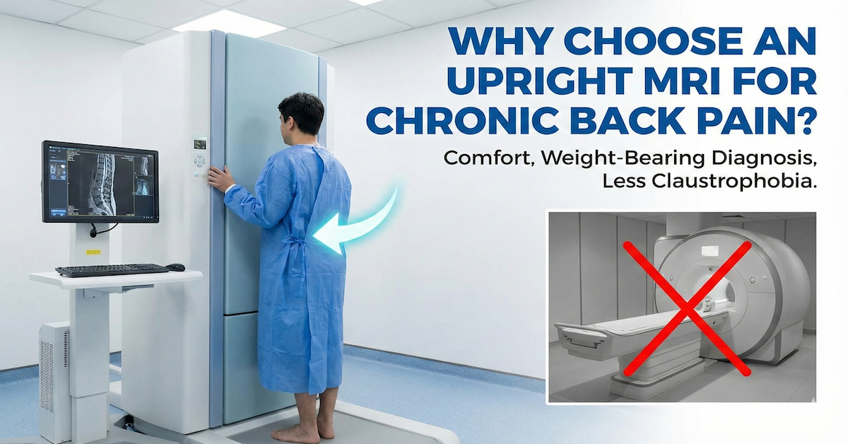
What Upright Foraminal Imaging Looks Like in Practice
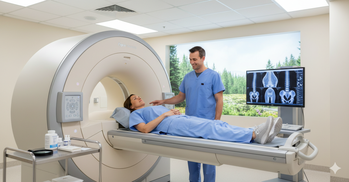
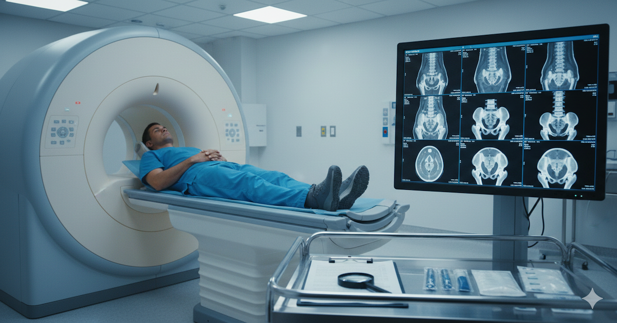
The practical experience of an upright MRI for foraminal evaluation is straightforward. The patient sits in an open scanner that allows imaging in the seated position. For cervical spine evaluation, different head positions can be captured. For lumbar spine evaluation, both neutral and flexed or extended positions are available. There is no enclosed tunnel, and the majority of patients find the experience significantly more comfortable than a conventional scanner.
The images are read by a radiologist familiar with the positional and weight-bearing context of the scan. More information on what is included in a lumbar or cervical evaluation is available on the back and spine scan page at Upright MRI of Deerfield.
Frequently Asked Questions
What is foraminal narrowing and how does it cause pain?
Foraminal narrowing, also called foraminal stenosis, is a reduction in the size of the openings through which nerve roots exit the spinal column. When these openings become too small, the nerve root inside can be compressed, causing pain, tingling, numbness, or weakness that radiates along the nerve's path into the arm or leg.
Can foraminal narrowing be missed on a standard lying-down MRI?
Yes, and this is relatively common. Because the foramina can partially open when spinal load is removed, a lying-down MRI may show only mild narrowing in a patient who has significant nerve compression when standing or sitting. Upright imaging captures the true loaded dimensions of the foramen.
Is upright MRI the same as open MRI?
Not exactly. Open MRI typically refers to a scanner with an open design that reduces claustrophobia but still images the patient lying down. Upright MRI specifically allows the patient to be scanned in a seated or standing position, which is the key diagnostic advantage for weight-bearing and positional conditions.
How do I know if my symptoms are caused by foraminal narrowing?
Foraminal narrowing typically produces radiating symptoms that follow a specific nerve root pattern, such as pain and numbness down one arm or leg. Symptoms often worsen with certain positions, particularly neck extension or lumbar extension. A positional MRI combined with a clinical examination is the most reliable way to confirm the diagnosis.
Do I need a referral from my doctor to get an upright MRI?
Many upright MRI facilities accept self-referrals, meaning patients can schedule independently without a physician referral. The resulting scan and radiologist report can then be shared with your doctor, specialist, or surgeon to inform further treatment decisions.
The Bottom Line
Foraminal narrowing is a condition where the position of the spine during imaging makes a real difference to what is found. An upright MRI images the spine under the gravitational load that actually compresses these nerve openings, providing a more accurate and clinically relevant picture than a lying-down scan can offer.
Upright MRI of Deerfield provides positional MRI imaging specifically designed to capture the spine as it functions in real life. If you have been told your scan is normal but your symptoms say otherwise, or if foraminal narrowing is suspected but not yet confirmed, the team at Upright MRI of Deerfield is ready to help you get the clearer picture you need.
SHARE THIS POST:
Leave a Comment:

The World's Most Patient-Friendly MRI. A comfortable, stress-free, and completely reliable MRI scan. We offer patients an open, upright, standup MRI experience that helps those who are claustrophobic and stress being in a confined area. Upright MRI of Deerfield is recognized as the world leader in open MRI innovation,
Our Recent Post


READ PATIENT TESTIMONIALS
Upright MRI of Deerfield.
Susan D.,
Highland Park, 39
I am going to tell everyone about your office! This was a great experience after I panicked in other MRI machines and had to leave. Thank you so much.

Judith B.,
Milwaukee, 61
I suffer from vertigo and other MRIs do not work. This was wonderful…absolutely NO discomfort at all. The MRI was so fast…I wanted to stay and watch the movie! Mumtaz was great. His humor really put me at ease. I’ve already recommended Upright MRI to friends.
Delores P.,
Glencoe, 55
Everything is so nice and professional with your place. I have been there a couple of times. My husband and I would not go anywhere else.

