
When Is a Knee MRI Needed in the Upright Position?
The knee is one of the most commonly injured joints in the body, and MRI is the standard tool for evaluating meniscal tears, ligament injuries, cartilage damage, and bone pathology before any surgical decision is made. For most acute injuries, a supine knee MRI performed in a conventional closed-bore scanner provides excellent diagnostic information and is entirely appropriate. But there is a meaningful subset of knee complaints where lying flat with the knee extended or slightly flexed does not reproduce the mechanics that produce symptoms, and where the information available from that position is genuinely incomplete.
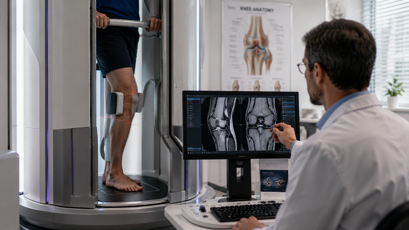
The knee is a weight-bearing joint whose function is defined by what happens when a person stands, walks, and bends under gravitational load. The way the menisci compress and distribute force, the way cartilage deforms and recovers, the way ligament tension changes with loading: all of these depend on body weight being transmitted through the joint in the way it actually is during daily activity. A scan taken while the patient is unloaded and horizontal captures a different set of conditions from the ones that produce most knee symptoms.
Quick Answer: An upright knee MRI is most clinically valuable when the patient's symptoms are clearly activity-dependent or weight-bearing-dependent, when a supine scan has been inconclusive or does not explain the clinical picture, when meniscal extrusion or cartilage deformation under load is suspected, or when subtle ligamentous laxity and patellofemoral malalignment are better assessed in the standing position. The upright position allows body weight to act through the knee during imaging, which reveals load-dependent pathology invisible in the supine state.
What Changes in the Knee When You Stand
The transformation of the knee from its supine to standing state involves multiple simultaneous changes that affect the appearance and behaviour of virtually every structure in the joint. The tibiofemoral compartments compress under load, reducing the joint space and compressing the menisci. The menisci, which function as shock absorbers, flatten and spread under this compression. Articular cartilage deforms as the load passes through it. The position of the patella relative to the trochlear groove shifts as the quadriceps engage to stabilise the joint.
These changes are not minor. Studies comparing supine and weight-bearing knee imaging have consistently demonstrated that joint space measurements, meniscal dimensions, cartilage thickness at load-bearing points, and patellar position all differ meaningfully between the two states. For a joint whose pathology is fundamentally linked to what happens under load, these differences have direct clinical significance.
Meniscal Pathology and Load-Dependent Findings
The menisci are the structures most commonly associated with knee pain and surgical intervention in the adult population, and also the structures whose behaviour under load differs most from their appearance at rest. In the supine position, the menisci are partially decompressed and may sit closer to their normal anatomical position than they do during weight-bearing.
Meniscal extrusion, in which the meniscus shifts laterally beyond the tibial margin under load, is a finding associated with accelerated cartilage wear and early osteoarthritis progression. Extrusion that is minimal or absent in the supine position may become clinically significant in the standing scan. For patients with medial knee pain and a supine scan showing only mild meniscal degeneration, upright imaging that demonstrates significant extrusion under load provides a more complete picture of what is actually happening in the joint during daily activity.
Meniscal tears that are mobile, meaning that the torn fragment shifts with joint loading and movement, may also present differently in the upright position. A tear whose fragment displaces into the joint line under weight-bearing produces both more pain and a more apparent MRI finding when imaged in the position that causes the displacement.
Articular Cartilage: The Importance of Load
Articular cartilage evaluation is one of the most important applications of knee MRI and one where the imaging position has meaningful implications. Cartilage under load is structurally different from cartilage at rest: it deforms as the joint compresses, and areas of thinning, softening, or focal damage become more apparent when stress is passing through them.
The lateral and medial weight-bearing surfaces of the femoral condyles and the tibial plateau are the areas where load-bearing cartilage assessment is most clinically relevant, and these are precisely the areas where the difference between supine and upright imaging is greatest. Focal cartilage defects that are subtle on a resting scan can be more clearly delineated when the cartilage is being compressed in the position the patient adopts during the activities that produce their symptoms.
The principle behind weight-bearing MRI for cartilage evaluation is the same as for other joint structures: imaging the tissue in the condition it is in during the activities that cause symptoms provides more clinically relevant information than imaging it at rest.
Patellofemoral Disorders: A Position-Specific Problem
Patellofemoral pain syndrome and patellar instability are among the most common knee problems in younger and active patients, and both are fundamentally about the relationship between the patella and the trochlear groove during dynamic loading. The patella sits in the groove differently depending on the degree of knee flexion, the quadriceps activation level, and the load passing through the joint.
Conventional supine MRI with the knee in near-full extension captures patellar position in a state that may not reproduce the maltracking or instability that occurs during squatting, stair climbing, or running. An upright scan with the knee bearing weight and in a position of partial flexion that reproduces the patient's symptoms provides a significantly more relevant assessment of patellar tracking, tilt, and displacement.
Lateral patellar tilt and subluxation that are absent or minimal at rest can be clearly demonstrated during weight-bearing in the position that produces the patient's lateral retinacular tightness and anterior knee pain. This finding changes both the specificity of the diagnosis and the direction of subsequent management.
Ligamentous Laxity and Subtle Instability
Cruciate and collateral ligament tears with significant fibre disruption are well-visualised on supine MRI, and for complete tears the positional question is largely moot. The area where the limitations of supine imaging are most significant for ligament assessment is subtle or partial tears, chronic laxity without acute fibre disruption, and cases where the clinical concern is instability that manifests under functional loading rather than during static examination.
A knee that clinically demonstrates subtle anterior laxity with a positive Lachman test but returns an equivocal supine MRI may show more clearly demonstrable tibial translation under load in an upright scan. The ability to position the patient in a stance that applies the relevant stress to the ligament, rather than examining it under no load, provides a different type of information that complements rather than replaces the supine study.
Knee Osteoarthritis: Grading Under Real Conditions
The grading of knee osteoarthritis on MRI, which assesses the degree of cartilage loss, bone marrow oedema, osteophyte formation, and joint space narrowing, is typically performed on supine imaging. However, the functional impact of osteoarthritis on a patient's daily life is determined by what happens when they stand and walk, not by the static appearance of the joint at rest.
Joint space in the medial compartment, which is the most commonly narrowed compartment in knee osteoarthritis, is consistently reduced in the upright position compared to the supine position. This difference affects grading decisions and can influence whether a patient meets the threshold for surgical consultation or specific interventions. Patients whose supine scan suggests mild to moderate disease may show more significant load-bearing joint space narrowing that better reflects their level of functional impairment.
Patients with chronic knee pain and a supine scan that understates their symptoms often find that chronic pain and upright MRI provides the structural explanation for what they have been experiencing, improving the alignment between the imaging findings and the clinical reality.
Which Patients Should Be Referred for Upright Knee Imaging
The range of conditions needing positional imaging in the knee includes patellofemoral disorders, meniscal extrusion, weight-bearing cartilage assessment, chronic knee pain with inconclusive supine imaging, and cases of suspected subtle ligamentous laxity where load-dependent translation is the clinical concern.
Patients who describe pain specifically during weight-bearing activities and whose supine MRI has not provided a satisfying explanation for their symptoms are the strongest candidates. Athletes with activity-related knee pain that does not reproduce during the scan examination are another group where upright imaging adds meaningful information.
The positional MRI for joints approach is particularly valuable in the knee because the joint's primary function is weight-bearing, and imaging it in a non-weight-bearing state inevitably captures a physiologically different condition.
Clinicians wishing to refer patients for upright knee imaging will find detailed protocol and referral information available under physician referral protocols at Upright MRI of Deerfield, including guidance on how to specify the positions and loading conditions most relevant to each clinical scenario.
Frequently Asked Questions
Is an upright knee MRI more accurate than a standard MRI?
Neither is universally more accurate. They capture different states of the same joint. A supine scan is more accurate for detecting certain acute structural injuries. An upright scan is more accurate for assessing load-dependent changes in meniscal position, cartilage under compression, patellar tracking, and joint space narrowing under body weight. The choice depends on the clinical question.
Can children have upright knee MRI?
Yes. Upright MRI is suitable for patients of all ages who can cooperate with the positioning requirements. For very young children who cannot remain still for the duration of the scan, a conventional scanner may be more practical. For older children and adolescents with activity-related knee pain, upright imaging can be very informative.
How long does an upright knee MRI take?
A standard upright knee MRI typically takes between 30 and 45 minutes. If multiple positions or loading conditions are being imaged, the time may be slightly longer. The open design of upright MRI systems tends to make the experience more comfortable than a conventional closed-bore scanner, particularly for patients with claustrophobia.
Will I need to bear full body weight during an upright knee MRI?
The degree of weight-bearing during the scan depends on the clinical protocol. Some studies involve full standing weight-bearing; others involve partial loading with the knee in a specific position. The imaging team will position you in the way most likely to capture the clinically relevant information for your specific situation.
Can an upright knee MRI be done if I have already had a supine MRI?
Yes. An upright study can be performed as a follow-up to an inconclusive or negative supine scan when the clinical picture suggests positional or load-dependent pathology. The upright scan can also be compared directly to the previous supine study to identify changes in joint space, meniscal position, or patellar tracking that appear under load.
The Bottom Line
A knee MRI in the upright position is clinically indicated when symptoms are weight-bearing-dependent, when a supine scan has not explained the clinical picture, or when the specific pathology being evaluated, meniscal extrusion, patellofemoral maltracking, load-bearing cartilage changes, or subtle ligamentous laxity, is best assessed under functional load.
Upright MRI of Deerfield provides weight-bearing knee imaging for patients in the Chicago area and Midwest. If your knee symptoms have not been fully explained by conventional imaging, a consultation or referral for positional imaging is a practical next step.
SHARE THIS POST:
Leave a Comment:

The World's Most Patient-Friendly MRI. A comfortable, stress-free, and completely reliable MRI scan. We offer patients an open, upright, standup MRI experience that helps those who are claustrophobic and stress being in a confined area. Upright MRI of Deerfield is recognized as the world leader in open MRI innovation,
Our Recent Post
READ PATIENT TESTIMONIALS
Upright MRI of Deerfield.
Susan D.,
Highland Park, 39
I am going to tell everyone about your office! This was a great experience after I panicked in other MRI machines and had to leave. Thank you so much.

Judith B.,
Milwaukee, 61
I suffer from vertigo and other MRIs do not work. This was wonderful…absolutely NO discomfort at all. The MRI was so fast…I wanted to stay and watch the movie! Mumtaz was great. His humor really put me at ease. I’ve already recommended Upright MRI to friends.
Delores P.,
Glencoe, 55
Everything is so nice and professional with your place. I have been there a couple of times. My husband and I would not go anywhere else.