
Is an Upright MRI More Accurate for Degenerative Disc Disease?
Degenerative disc disease is a common description for age related or wear related changes in the discs of the spine. Those changes can include loss of water content, reduced disc height, small tears, bulging, and changes in the joints or bone around the disc. The amount seen on a scan does not always match the amount of pain a person feels.
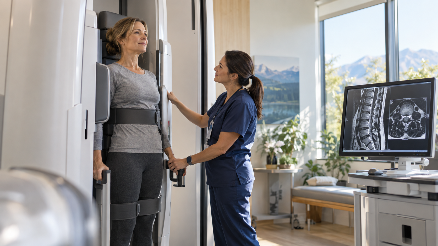
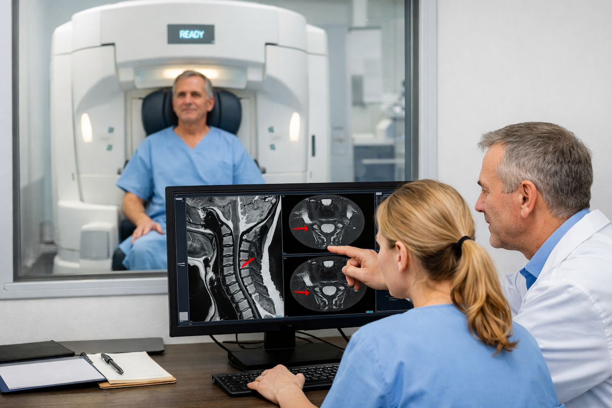
That mismatch leads many patients to ask whether the position of the MRI matters. A conventional scan shows the spine while the patient lies down, but symptoms from disc degeneration often become more noticeable during sitting, standing, walking, or bending. Upright imaging examines the spine while it is carrying more of its normal load.
Quick Answer: An upright MRI can be more accurate for showing how degenerative disc disease behaves under body weight, especially when symptoms change with posture. It can reveal changes in disc height, bulging, alignment, or nerve space that look less pronounced while lying down. It is not more accurate for every finding, so the best MRI position depends on the symptoms and the clinical question.
What Does Degenerative Disc Disease Mean on MRI?
The name can sound alarming, but degenerative disc disease is not an infection or a condition that always keeps getting worse. It refers to structural changes in one or more spinal discs. Some are part of normal aging, while injury, repeated strain, genetics, smoking, and other factors can influence how early or how strongly they appear.
A back and spine MRI examination may show darker disc signal from reduced water content, loss of disc height, bulging or herniation, endplate changes, and narrowing around the nerves. The report may also describe facet joint arthritis or thickened ligaments because disc degeneration often affects more than the disc alone.
These findings need clinical context. A person can have obvious degeneration with little pain, while another person has serious symptoms from a relatively subtle change in a critical location. The goal is to find the abnormality that best matches the symptom pattern.
Why Can Upright Imaging Change the Assessment?
Standing and sitting place different forces across the spine than lying down. Under load, a worn disc may lose additional height, a bulge may become more prominent, and the openings around the nerves may become smaller. Spinal alignment can also change when muscles, joints, and ligaments respond to gravity.
This is why weight bearing MRI can improve spine assessment for carefully selected patients. It adds information about function and position instead of replacing the detailed anatomical information that MRI already provides.
Research comparing standing and loaded scans has documented measurable changes in disc shape, disc height, spinal curvature, canal dimensions, and foraminal space. Those changes are real, but their importance depends on whether they explain the patient's pain, numbness, weakness, or limits in daily activity.
Is Upright MRI Always More Accurate?
No. Accuracy depends on what the clinician needs to know. A conventional MRI may provide excellent detail for a fixed disc herniation, tumor, infection, fracture, inflammation, or other finding that does not depend on posture. A higher field scanner may also offer advantages for certain fine anatomical details.
Upright MRI is strongest when the question itself involves loading or movement. The range of conditions assessed with positional MRI includes disc degeneration, spinal stenosis, spondylolisthesis, and other problems that can change between lying, sitting, and standing.
The most accurate examination is therefore the one with the right body area, sequence, position, and image quality for the suspected problem. The referral should make that problem clear so the imaging protocol can be chosen around it.
Which Degenerative Changes May Look Different Upright?
Disc degeneration can affect several connected structures. Upright imaging may show a different degree of change in one area while another finding remains stable.
· Disc height may decrease further under load, especially at a worn segment.
- A broad bulge or focal protrusion may change in contour or relation to a nerve.
- The neural foramina may narrow as disc height and spinal alignment change.
- A vertebra may shift relative to the one below it, revealing positional instability.
- The central spinal canal may become smaller in a posture that reproduces symptoms.
One clinically important example is foraminal narrowing seen on upright MRI. If a degenerating disc loses height, the opening for the nerve can become tighter, particularly in a loaded or extended position.
How Do Flexion and Extension Improve the Comparison?
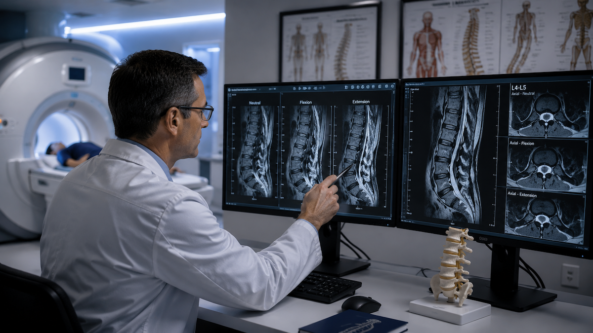
Neutral upright images show the spine under load without deliberately bending it. Flexion and extension add controlled forward and backward positions. Comparing them can show whether a segment is stable and whether the available space for nerves changes with movement.
The difference between neutral and flexion MRI is useful when symptoms follow a repeatable movement. A person whose pain increases while leaning backward may need a different positional comparison from someone whose symptoms appear during prolonged sitting or forward bending.
Movement based images can also show that a degenerative finding is stable. That can be just as useful as finding abnormal motion because it prevents the report from assuming instability based only on disc wear.
When Might an Upright Degenerative Disc Disease MRI Help Most?
The examination is most likely to add value when there is a clear reason to suspect that gravity or posture changes the problem. A doctor may consider it after reviewing the symptom pattern, examination, and any earlier imaging.
- Pain, numbness, or weakness becomes worse during standing, walking, or sitting.
- Symptoms improve substantially when lying down.
- A prior supine MRI shows mild degeneration that does not explain significant symptoms.
- The clinician suspects dynamic narrowing or movement at a degenerative spinal level.
- Surgical or procedural planning requires a better view of which level changes under load.
For patients with persistent symptoms and limited answers, the role of upright MRI in chronic back pain is to test a specific mechanical explanation. It should not be ordered only because degeneration appears on an older scan.
Can Upright MRI Improve Treatment or Surgical Planning?
Imaging does not choose a treatment by itself, but it can change the information available to the treating clinician. If a level narrows or shifts only under load, that may affect which structure is considered responsible for symptoms and which treatment options deserve discussion.
A dynamic spine MRI for surgical planning can help show whether more than one level changes with position or whether an apparent problem stays stable. Surgeons still weigh the examination, symptom severity, response to nonoperative care, and the risks and benefits of any procedure.
For nonsurgical care, a better match between symptoms and imaging may help guide physical therapy, activity changes, injections, or further testing. It may also show that the degenerative findings are unlikely to be the main source of pain, which can redirect the evaluation.
What Are the Limits of Upright MRI for Disc Degeneration?
The scan has practical and diagnostic limits. Upright scanners and protocols vary, and image detail can differ from a conventional high field MRI. Movement during the scan may reduce image quality, while pain can make it difficult to hold the requested position long enough.
Position dependent changes are not automatically the cause of symptoms. Degenerative findings are common, especially with age, so the report must avoid treating every visible change as a diagnosis. The size of a bulge or the grade of disc wear does not measure pain.
A normal supine scan also should not be dismissed. When symptoms are clearly positional, flexion imaging after a normal supine MRI can answer a focused follow up question, but it is not a routine requirement for everyone with back pain.
How Should You Prepare for an Upright Spine MRI?
Preparation is similar to other MRI examinations. The imaging team will screen for implanted devices, metal, prior procedures, and any other safety concerns. You may be asked to remove jewelry, watches, hearing aids, removable dental items, and clothing with metal parts.
Tell the technologist which positions bring on symptoms and whether you can safely hold them. The team may obtain neutral images first and then add a seated, standing, flexed, or extended position based on the prescription and protocol.
Knowing what happens during an MRI scan can make the appointment easier. You will need to remain still during each sequence, and you should speak up if pain, weakness, or anxiety makes a position hard to maintain.
How Should the Results Be Interpreted?
The radiologist may describe disc signal, height, contour, alignment, canal size, and foraminal size in one or more positions. The important question is whether the loaded images add a meaningful finding that corresponds to the patient's symptoms.
Results should be reviewed with the referring or treating clinician. That conversation can separate common age related findings from changes that are likely to matter, clarify whether more testing is needed, and connect the report to a realistic care plan.
Seek urgent assessment for new bladder or bowel control problems, numbness around the saddle area, rapidly worsening weakness, fever with severe back pain, or major trauma. These warning signs require prompt medical attention rather than routine follow up.
Frequently Asked Questions
Does degenerative disc disease show better on an upright MRI?
It can show load dependent effects of disc degeneration more clearly, including additional loss of height, a change in bulging, or reduced nerve space. Fixed degenerative changes may already be shown well on a standard MRI.
Can an upright MRI show how severe disc degeneration is?
It can show how the affected level behaves in selected positions, which adds useful information about function. Severity still depends on the full imaging pattern and how well it matches symptoms, not on one measurement alone.
Is degenerative disc disease always painful?
No. Many people have degenerative changes on MRI without significant pain. A clinician looks for a close match between the image, the location of symptoms, and physical findings before linking degeneration to pain.
Should I repeat a normal MRI in an upright scanner?
A repeat scan may be reasonable when symptoms are persistent, clearly position dependent, and not explained by the earlier examination. The referring clinician should decide whether a positional study is likely to answer a specific unresolved question.
Can upright MRI replace a conventional MRI?
Sometimes it can provide the needed examination, but it is not a universal replacement. The right choice depends on the suspected condition, required image detail, available protocol, safety factors, and the positions the patient can tolerate.
Conclusion
An upright MRI can give a more accurate picture of how degenerative disc disease behaves under body weight, especially when symptoms change with posture. It is most useful as a targeted examination, and its findings must be interpreted alongside the clinical history and physical examination.
Upright MRI of Deerfield offers positional spine imaging that can compare the back in relevant seated, standing, or movement based positions. Patients can visit the website or contact the practice to ask about prescriptions, insurance, and scheduling.
SHARE THIS POST:
Leave a Comment:

The World's Most Patient-Friendly MRI. A comfortable, stress-free, and completely reliable MRI scan. We offer patients an open, upright, standup MRI experience that helps those who are claustrophobic and stress being in a confined area. Upright MRI of Deerfield is recognized as the world leader in open MRI innovation,
Our Recent Post


READ PATIENT TESTIMONIALS
Upright MRI of Deerfield.
Susan D.,
Highland Park, 39
I am going to tell everyone about your office! This was a great experience after I panicked in other MRI machines and had to leave. Thank you so much.

Judith B.,
Milwaukee, 61
I suffer from vertigo and other MRIs do not work. This was wonderful…absolutely NO discomfort at all. The MRI was so fast…I wanted to stay and watch the movie! Mumtaz was great. His humor really put me at ease. I’ve already recommended Upright MRI to friends.
Delores P.,
Glencoe, 55
Everything is so nice and professional with your place. I have been there a couple of times. My husband and I would not go anywhere else.