
What Types of Cancer Do Doctors Detect With MRIs?
MRIs are incredibly useful and important tools for cancer detection. In this article, we will explore MRIs, how they work, what you should know before you get one, and, most importantly, the types of cancers they detect.
What Is an MRI?
Before we can explore what types of cancer doctors detect with MRIs, it’s helpful to understand the procedure. MRI stands for magnetic resonance imaging. As the word “imaging” suggests, MRIs are tools doctors use to observe, or “image,” the inside of a human body. Using an MRI, doctors can detect cancers, which will be the focus of this article, but they can also detect other ailments, like:
- Spinal cord injuries
- Brain injuries
- Multiple Sclerosis, an auto-immune disease
- Eye problems
- Brain infections
- Damage to the optic nerve
- And more
MRIs are life-saving devices because, when properly utilized, doctors can leverage the technology to find diseases, especially cancers, before they spread and further damage the body. Doctors use the information gathered from MRIs to create medical plans. Patients also return to the devices during their treatments to see the progression of their diseases.
How Does an MRI Work?
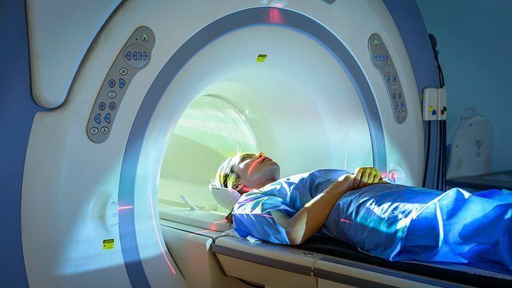
Even if you do not have firsthand experience with MRI machines, you have probably seen one in movies or television. They are large cylindrical tubes that contain a very strong magnet. To use an MRI machine, patients lie on a plank that lifts and travels into the machine, surrounding the patient with the intense magnetic field. The machine sends radio frequencies through the person’s body. The radiofrequency waves detect signals from the nuclei of hydrogen atoms inside the patient. The information gathered from these signals travels to a computer, which converts them into black and white images.
Doctors may also inject contrast materials into the patient’s body in order to produce clearer images from the MRI. Contrast, after the body absorbs it, increases the speed at which tissues respond to radio and magnetic waves.
After an MRI, patients can see cross-sectional images of their internal organs. It’s helpful to think of an MRI as a camera that takes images from several angles. Using an MRI, you can simultaneously look at your body from the side, front, or even above your head. MRIs show images of soft tissues that are difficult to see with other imaging tests.
Other Medical Imaging Technologies
Of course, MRIs are not the only tools doctors use to analyze a patient’s insides. For many years, doctors have leveraged several technologies to uncover what goes on under the skin. In this section, we will explore a few other medical imaging technologies you should know.
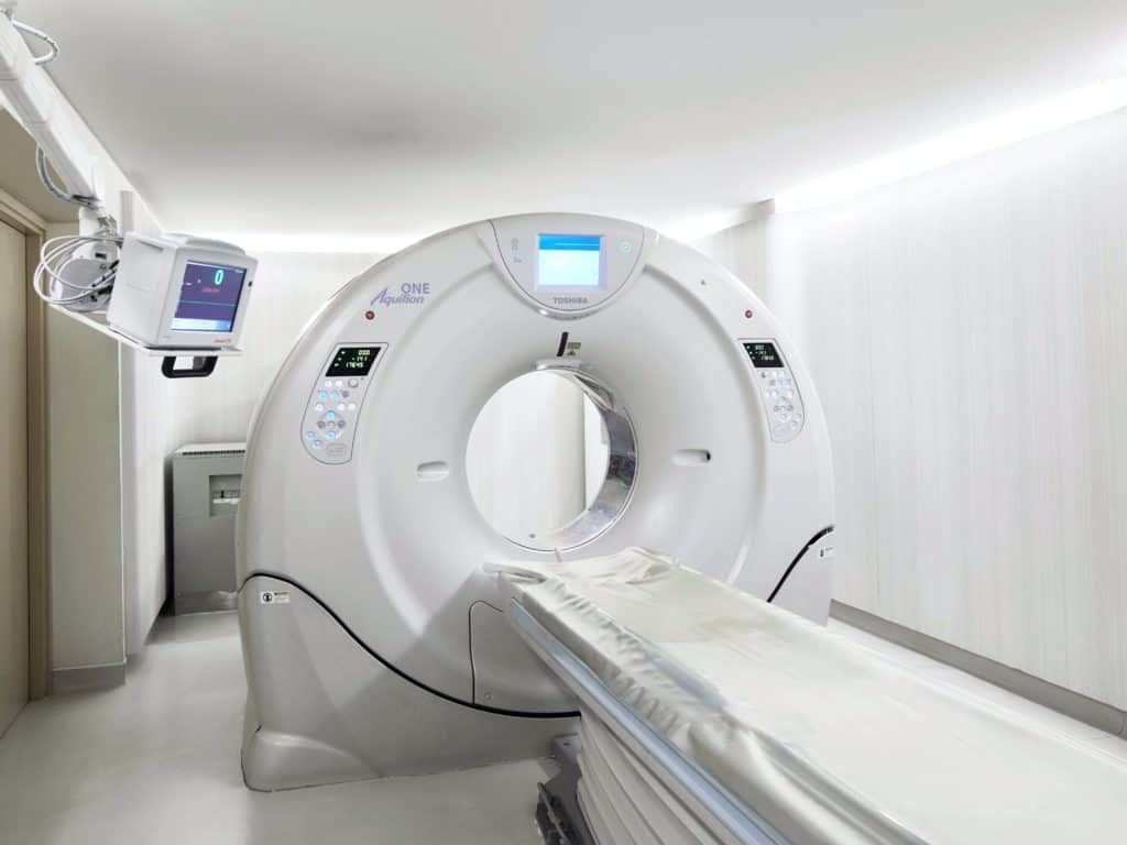
Computed Tomography
Computed topography, which we often abbreviate as a CT scan, is medical imaging technology that is similar to an MRI but different in several vital ways. Like an MRI, CT scans lift people into a tube to analyze their bodies. However, CT scans use a more traditional X-ray system to create its images. A CT scan takes multiple pictures in quick secession to develop the cross-sectional images doctors need. They tend to be more detailed than plain X-rays. Doctors may use CT scans to find tumors, diseases, and accident-related injuries.
Sonography
Also known as ultrasound imaging, sonography is a medical imaging tool that utilizes sound waves. To administer an ultrasound, doctors apply gel on the spot where they want to develop an image. They then put a probe, which is called a transducer, on the spot. The transducer creates an electrical signal that gets converted into images. Usually, doctors turn to sonography when they want to look at the growth of a fetus. But sonography also comes in handy when doctors want to examine symptoms involving infection, pain, or swelling. Using sonography, doctors can look at the gallbladder, liver, kidney, brain, spine, heart, and other organs.
Vascular Interventional Radiography
Doctors conduct vascular interventional radiology techniques usually by putting a needle through an incision and into a person’s skin. Sometimes, doctors use wires or catheter tubes. This procedure uses several imaging methods, including tomography, X-ray fluoroscopy, and ultrasounds to make pictures of a person’s body.
What Types of Cancers Do Doctors Detect With MRIs?
We have already explored several ailments MRIs detect. But now we focus on their application for one of the most dangerous types of diseases: cancer. Nearly 2,000,000 people a year receive a cancer diagnose in the United States. Being diagnosed with cancer is a life-altering event. Many die from cancers they could have treated had they found the tumor earlier, which is why it’s so important to check for cancer regularly. Doctors can detect several types of cancers with MRIs.
MRIs, especially MRIs with contrast dye, are excellent at detecting cancers on the brain and spinal cord. The functional MRI of the brain, or the fMRI, is a type of MRI that specially studies the brain’s anatomy. Using MRIs, doctors can see if the cancer metastasized or spread throughout the body. Doctors utilize the information from MRIs to make plans for surgery or radiation therapy, two of the best practices for fighting against cancer.
What Should I Know Before I Get an MRI?
Although an MRI can be intimidating, it is a painless process for most people, though some experience nausea and anxiety. There is little to no special preparation for an MRI. Doctors do not require patients to follow any dietary rules before the procedure. Before an MRI, patients must make their doctors aware of metal in their bodies. They must remove all the metal they can and wear a gown.
Patients lie on a flat table while technicians operate the machine. Patients may feel a warm sensation on the part being analyzed. The machine makes louds noises which some describe as a thumping, whirring, or clicking. Thankfully, technicians usually give patients earplugs that pipe in music to block out the sound. A scan could take anywhere between 45 and 60 minutes.
Conclusion
In this article, we explored important information that patients need to know about MRIs and cancer. We talked about the different cancers that MRIs detect best, as well as how to prepare for an MRI. For those about to undergo an MRI, hopefully this article cleared up some of the confusion.New Paragraph
SHARE THIS POST:
Leave a Comment:

The World's Most Patient-Friendly MRI. A comfortable, stress-free, and completely reliable MRI scan. We offer patients an open, upright, standup MRI experience that helps those who are claustrophobic and stress being in a confined area. Upright MRI of Deerfield is recognized as the world leader in open MRI innovation,
Our Recent Post


READ PATIENT TESTIMONIALS
Upright MRI of Deerfield.
Susan D.,
Highland Park, 39
I am going to tell everyone about your office! This was a great experience after I panicked in other MRI machines and had to leave. Thank you so much.

Judith B.,
Milwaukee, 61
I suffer from vertigo and other MRIs do not work. This was wonderful…absolutely NO discomfort at all. The MRI was so fast…I wanted to stay and watch the movie! Mumtaz was great. His humor really put me at ease. I’ve already recommended Upright MRI to friends.
Delores P.,
Glencoe, 55
Everything is so nice and professional with your place. I have been there a couple of times. My husband and I would not go anywhere else.

