
Why Is a Supine MRI Not Always Enough for Diagnosing Spine and Joint Issues?
Magnetic resonance imaging, or MRI, is one of the most advanced tools doctors use to diagnose spine and joint conditions. It provides detailed views of muscles, nerves, and soft tissues without radiation exposure. However, traditional supine MRIs, where patients lie flat during the scan, don’t always tell the full story. For many people, pain or discomfort occurs only when standing, sitting, or moving, which means certain conditions can remain hidden when the body is relaxed. This article explains why a supine MRI isn’t always enough, how body position affects diagnosis, and when an Upright MRI may be a better option.

What Is a Supine MRI?
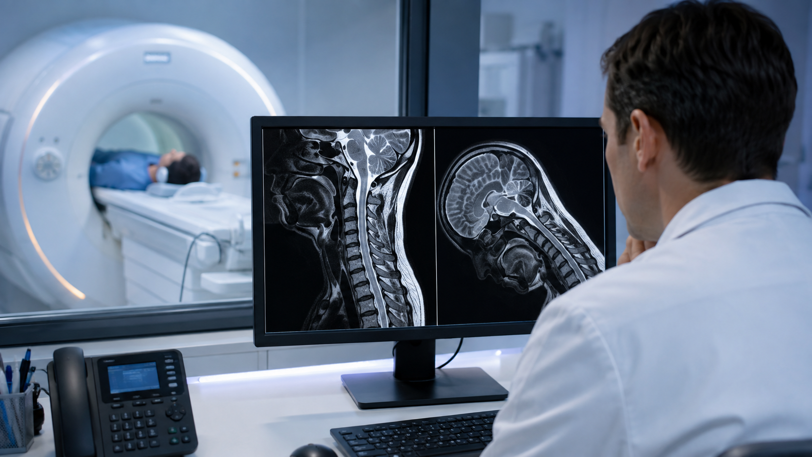
A supine MRI is the standard imaging method most people are familiar with. During the scan, you lie flat and still on a table that moves inside a cylindrical scanner. The machine captures detailed images of your internal structures, helping doctors evaluate the spine, joints, or soft tissues. Supine MRIs are excellent for showing clear images of muscles, discs, and ligaments, but they also come with a limitation gravity isn’t acting on your body the way it does when you’re upright. This means certain issues related to posture or movement might not be visible.
Why Spine and Joint Conditions Often Change with Body Position
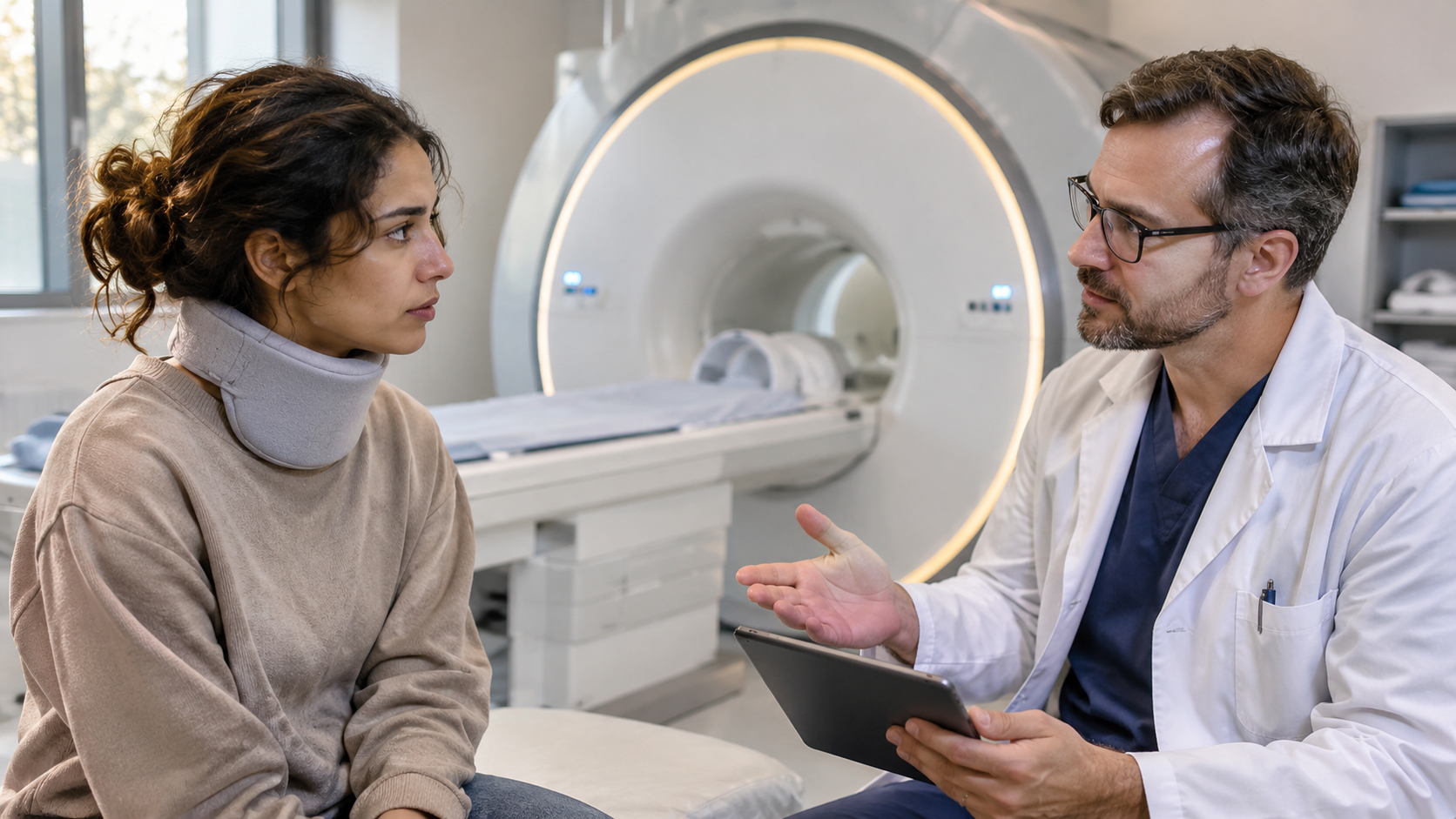
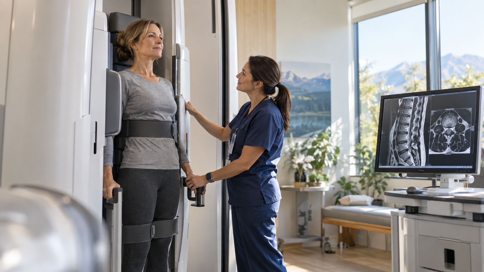
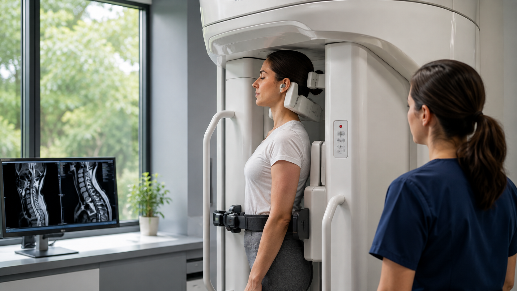
Our spines and joints behave differently under pressure. When you’re standing or sitting, gravity compresses your spine and joints, changing how discs, bones, and muscles align. For people with back or neck problems, this added pressure often triggers pain that disappears when lying down. A supine MRI doesn’t capture these real-world conditions. As a result, a patient might have normal results on a traditional MRI, even though pain persists in everyday life.
Conditions like herniated discs, spinal misalignment, and knee instability can shift position depending on posture. This is why imaging that reflects natural, weight-bearing positions like an Upright MRI can reveal more accurate diagnostic information.
Common Problems Missed in Supine MRIs
Several conditions are known to go undetected or appear less severe in traditional MRIs. These include:
- Herniated or bulging discs that flatten when lying down but press on nerves when standing
- Spinal stenosis, where the spinal canal narrows under gravity
- Joint instability that only occurs during movement or pressure
- Spondylolisthesis, when one vertebra slips forward, often more visible in upright scans
These conditions are responsible for much of the chronic back, neck, and joint pain patients experience. Missing them during a supine scan can lead to misdiagnosis or incomplete treatment plans.
The Importance of Functional Imaging for Spine and Joint Health
Functional imaging means evaluating the body in motion or under natural stress. Unlike static scans, functional or positional imaging captures the spine and joints as they function in real life. This makes a significant difference in understanding how pain develops and where it originates.
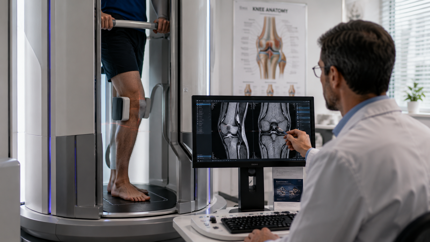
For example, a patient with knee pain may show no abnormalities in a supine MRI, but an Upright MRI can reveal joint compression that occurs while standing. Similarly, spinal compression or nerve pinching that happens under load can only be seen when the patient is upright. Functional imaging helps doctors connect symptoms to visible structural changes, leading to a more complete diagnosis.
Why Supine MRI May Lead to Incomplete or Misleading Diagnoses
Because a supine MRI removes gravitational pressure, it can sometimes show a “best-case scenario” of your spine and joints rather than how they perform under real conditions. This may cause doctors to underestimate the severity of your condition or miss it entirely. The result is frustration for patients who are told everything looks normal, even though their pain continues.
Incomplete imaging can also lead to unnecessary or ineffective treatments, as the true cause of discomfort remains hidden. This is especially true for chronic back or neck pain patients whose symptoms worsen when upright or moving.
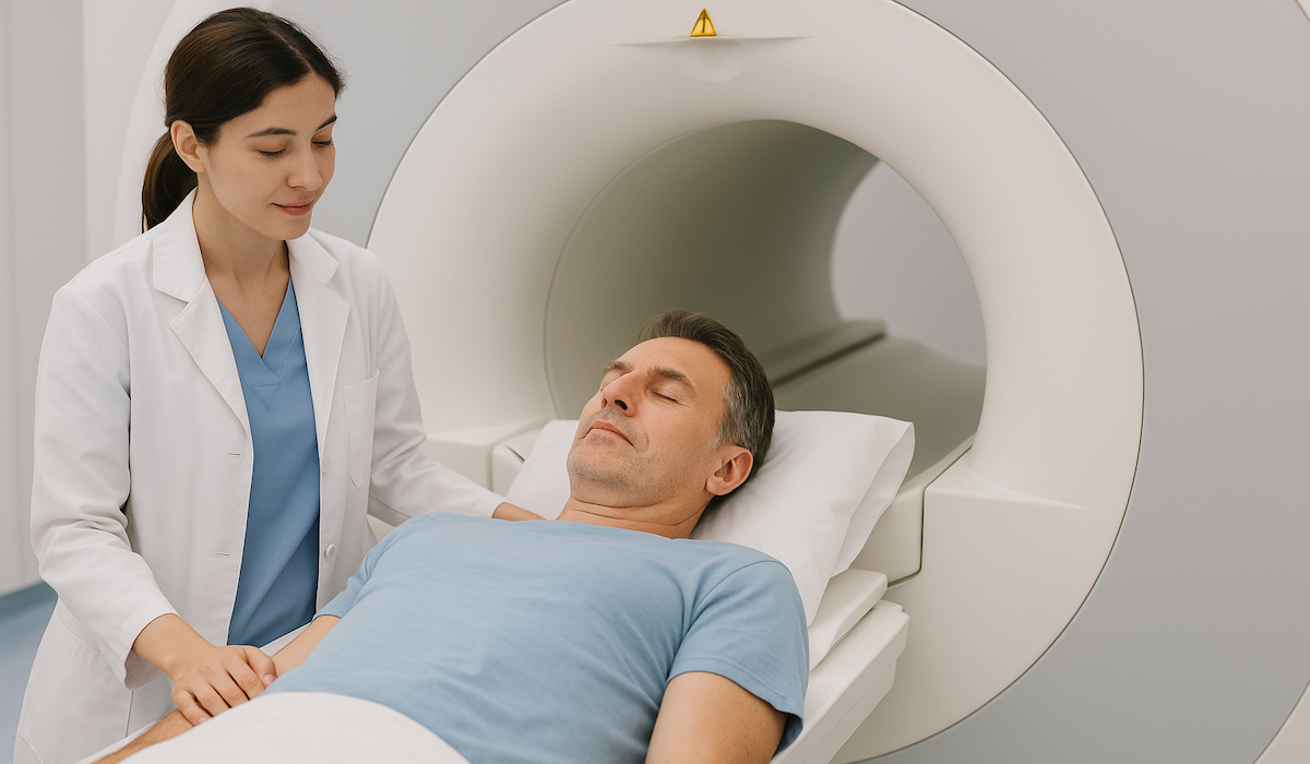
Comparing Supine MRI vs Upright MRI
Here’s a simple breakdown of how these two imaging techniques differ:
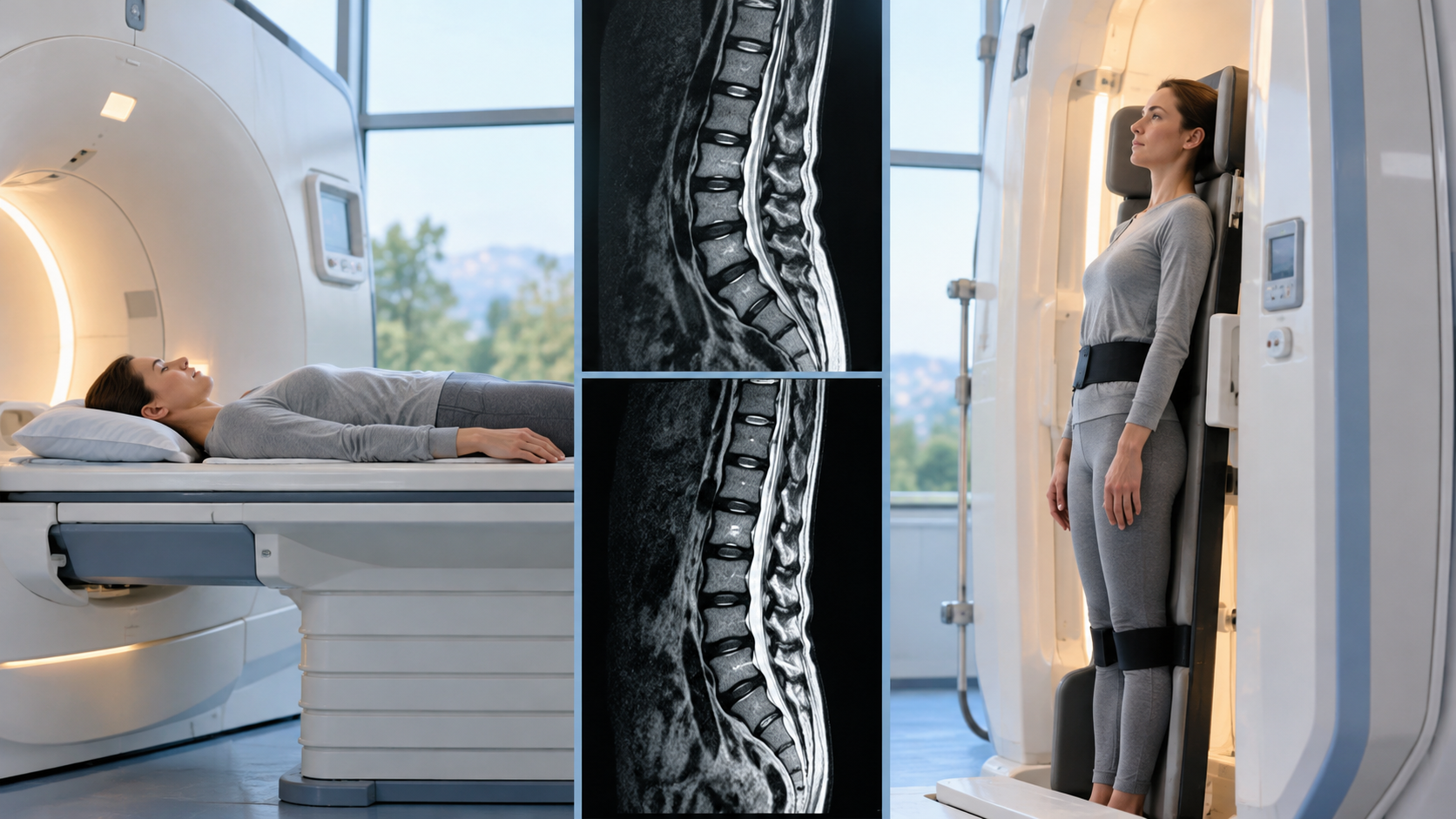
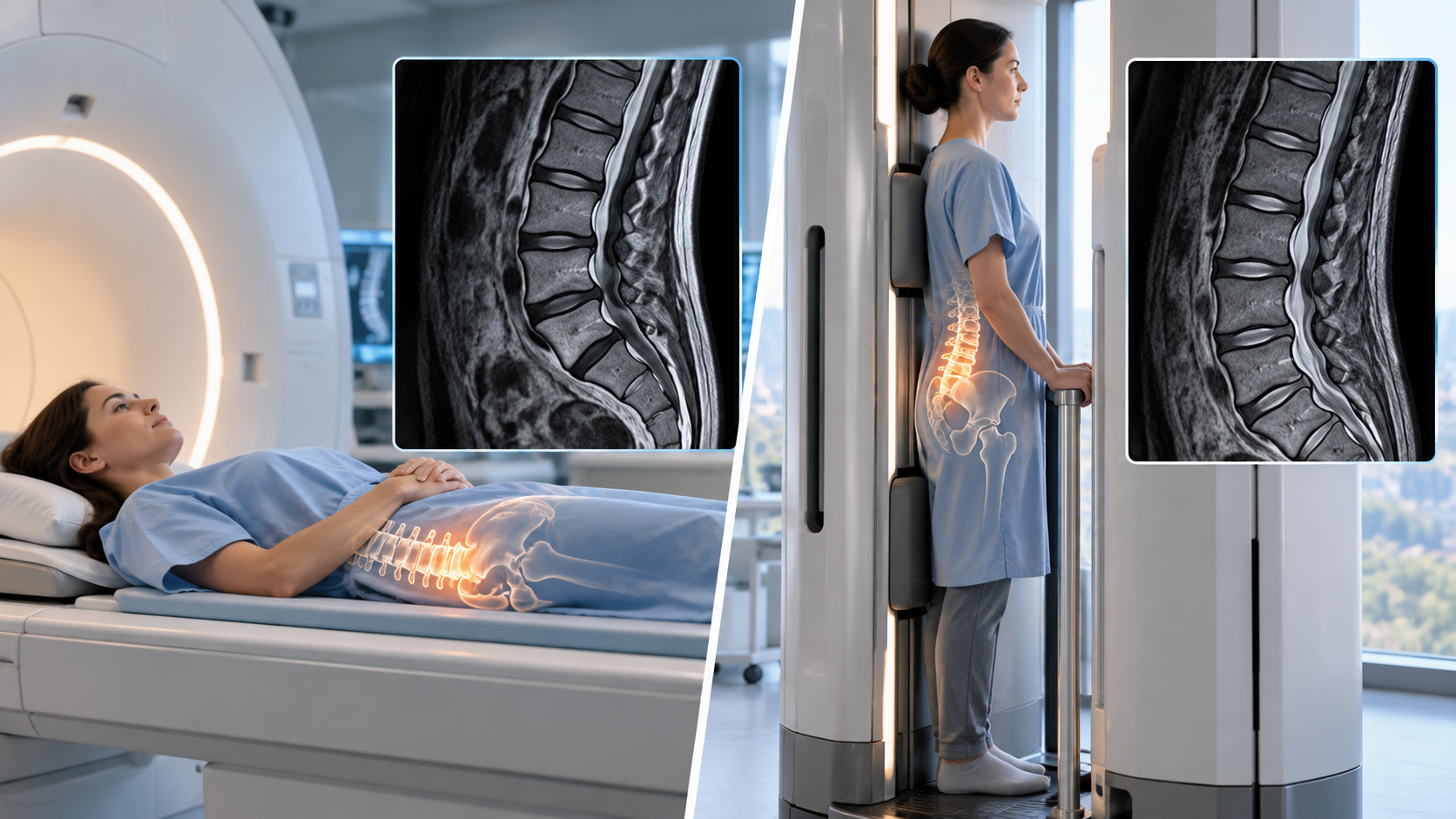
- Position: Supine MRI captures images with the patient lying flat; Upright MRI allows standing, sitting, or bending positions.
- Accuracy: Upright MRI can reveal weight-bearing problems that traditional MRIs miss.
- Comfort: Upright MRI’s open design reduces anxiety and claustrophobia.
- Use Cases: Supine MRI is ideal for static soft tissue imaging; Upright MRI is better for dynamic or posture-related pain.
Many doctors now recommend an Upright MRI when traditional imaging doesn’t match a patient’s symptoms.
When Should You Consider an Upright or Positional MRI Instead?
If your pain worsens when standing or sitting but disappears when lying down, an Upright MRI may be right for you. It’s also helpful if a previous MRI failed to identify the cause of your pain. Patients who experience joint instability, sciatica, or pressure-related discomfort often benefit the most. The open and comfortable design of Upright MRI machines also makes them a great option for people with claustrophobia or difficulty lying flat.
Conclusion
Supine MRIs remain a valuable tool in medical diagnostics, but they don’t always capture the full picture of how your body functions. For spine and joint issues that change with posture or movement, Upright MRI provides a more realistic and detailed view. This technology helps reveal hidden problems, leading to more accurate diagnoses and effective treatment plans.
If you’re struggling with persistent pain or inconclusive test results, UpRight MRI of Deerfield offers advanced Upright MRI technology designed to uncover what traditional scans might miss. Their patient-centered approach ensures a comfortable experience and clear, reliable results that help you and your doctor find the answers you need.
SHARE THIS POST:
Leave a Comment:

The World's Most Patient-Friendly MRI. A comfortable, stress-free, and completely reliable MRI scan. We offer patients an open, upright, standup MRI experience that helps those who are claustrophobic and stress being in a confined area. Upright MRI of Deerfield is recognized as the world leader in open MRI innovation,
Our Recent Post


READ PATIENT TESTIMONIALS
Upright MRI of Deerfield.
Susan D.,
Highland Park, 39
I am going to tell everyone about your office! This was a great experience after I panicked in other MRI machines and had to leave. Thank you so much.

Judith B.,
Milwaukee, 61
I suffer from vertigo and other MRIs do not work. This was wonderful…absolutely NO discomfort at all. The MRI was so fast…I wanted to stay and watch the movie! Mumtaz was great. His humor really put me at ease. I’ve already recommended Upright MRI to friends.
Delores P.,
Glencoe, 55
Everything is so nice and professional with your place. I have been there a couple of times. My husband and I would not go anywhere else.