
What Is Cranio-Cervical Instability and How Is It Detected
Brain, head, and spine conditions are some of the most painful and bothersome issues a person may face. Anyone who has had a concussion or contusion could tell how it negatively impacted their lives. One horrible, headache-inducing ailment you may not have heard of is cranio-cervical instability. CCI involves a corruption of the ligamentous connections. People with CCI experience looseness where the skull meets the spine. In this article on cranio-cervical instability and how it is detected, we delve into the details of this condition so that you can better understand how to spot it in yourself and others. Talk to a doctor if you think you might have CCI.
What Is Cranio-Cervical Instability?
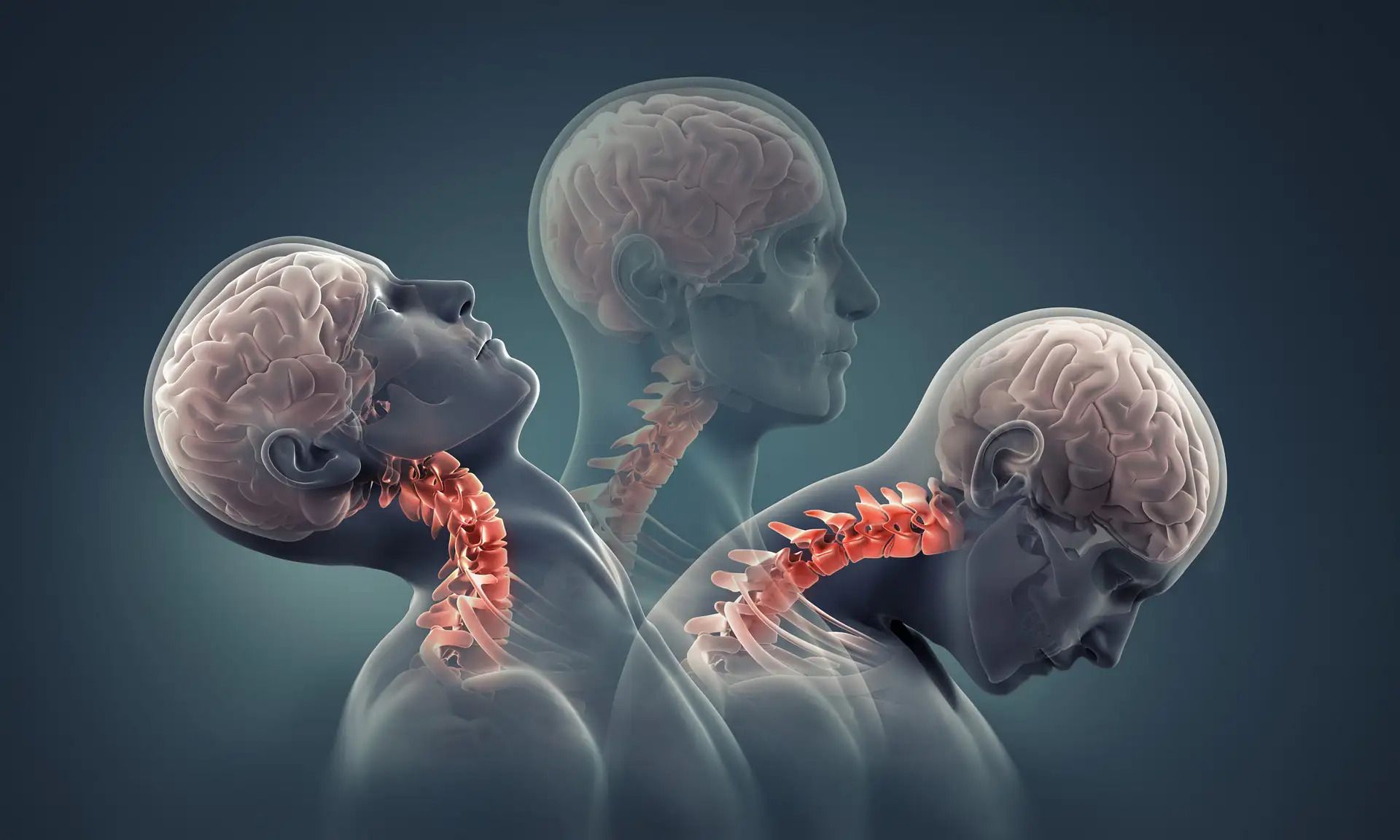
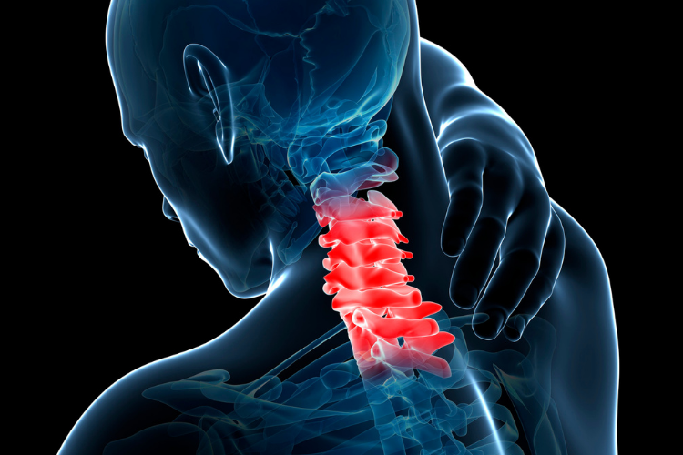
Also called the syndrome of occipitoatlantialaxial hypermobility, the condition cranio-cervical instability (CCI) can cause apathological deformations of the cerebellum, upper spinal cord, and brainstem. It is a structural instability of the craniocervical junction characterized by heightened mobility where the spine connects to the skull. People who suffer from CCI have stretched, ruptured, or weakened ligamentous connections. As a result, they experience a looseness where their neck connects to their head. Some people compare the feeling to being like a bobblehead. Normally, CCI evolves due to physical trauma like a car accident, congenital disorders like Down’s syndrome, or inflammatory diseases like arthritis. However, more recently, doctors have also reported seeing CCI in patients who live with connective tissue hereditary disorders like Ehlers-Danlos Syndrome.
Is CCI Serious and What Are Its Symptoms?
CCI may be a minor annoyance for some, but for others the symptoms can be very serious, even life-altering. A portion of people who have the condition are bed-bound. There are many symptoms attributed to the condition. Some of the most common symptoms include:
- Heavy headache: This is nothing like the occasional headaches most people encounter. A heavy headache entails a near-constant to constant headache in which the head feels like it weighs too much to be supported by the neck.
- Pressure headache: A pressure headache is sparked by a CSF flow impairment. It creates intracranial pressure that can be aggravated by “valsalva maneuvers” like laughing, coughing, sneezing, yawing, crying, or straining.
- Dysautonomia: This is a catch-all term for a number of disorders that involve an autonomic nervous system (ASN) that doesn’t work the way it should. People who suffer from dysautonomia grapple with:
o Heat intolerance
o Fainting
o Polydipsia (intense thirst)
o Chronic fatigue
o Tachycardia (rapid heart)
o Delayed gastric emptying
There are many other symptoms associated with craniocervical instability. These include:
- Pain in the neck
- Mixed or central sleep apnea
- Numbness or pain in the face
- Issues with balance
- Dizziness
- Vertigo
- Weak muscles
- Problems with vision
- Difficulty swallowing
- A reduced gag reflux
- Nausea
- Vomiting
- Irregular eye movements (known as downward nystagmus
- Paralysis
- And more
If you sustain the above symptoms and fear that you may have CCI, you should speak to a doctor as soon as possible to get a diagnosis. Working with a doctor, you can run tests to determine if you have this awful affliction.
How Is CCI Diagnosed?
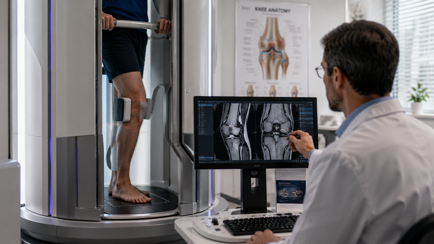
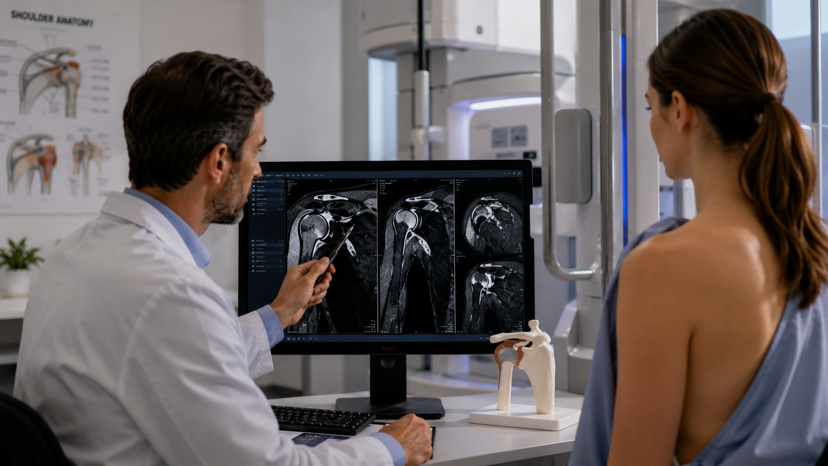
A CCI diagnosis requires the work of a medical professional. Doctors use a number of information sources to determine if a patient has CCI. They look at the symptoms exhibited, the patient’s medical history, and the results of medical imaging. For issues involving the brain, doctors prefer to test with magnetic resonance imaging (MRI), a medical imaging technique that utilizes a magnetic field and radio waves to develop detailed pictures of organs and tissues.
Neurosurgeons usually prefer to use the upright MRI machine, like the ones we provide at Upright MRI of Deerfield, to diagnose someone with CCI. Upright MRIs assess patients in weight-bearing or multiple positions. For example, it takes images of patients standing, sitting, or flexing. An upright MRI is the only imaging diagnostic tool that can see the damaged ligaments needed to diagnose a person with CCI. We frequently see patients recommended to us by attorneys and chiropractors.
Treatment
The conservative treatments for CCI include pain management, bracing the patient with a cervical collar, chiropractic treatment, and rest. However, these methods only offer limited relief. More experimental approaches may include stem cell therapy and prolotherapy.
When non-invasive approaches fail, surgery may be the best option. The surgery doctors use to treat CCI is called occipito-cervical fusion (OCF). The surgery attempts to stabilize the cranio-cervical junction. Usually, this entails the internal fixation of the skull to the spine through the use of mechanical screws and rods. Doctors use titanium hardware to fixate the occiput. They also utilize a rib graft, synthetic bone, or cadaver bone graft the fuse the bones.
Outcomes of Surgery
Little research about the outcomes of the surgery exists. While many enjoy improvements in dizziness, headaches, and frequent urination after the surgery, some deal with complications. The complications associated with the surgery can be intense. Common issues include wound infection, screw failure, and cerebrospinal fluid leakage. If the surgery goes poorly, some patients may need a second surgery to help with the infection or take out the hardware. OCF also entails a severe reduction in a person’s neck range of motion. However, for people who recover from CCI, this slight impairment is a small price to pay.
Conclusion
After reading this article, you hopefully better understand cranio-cervical instability and how is it detected. Cranio-cervical instability is a condition doctors are still trying to understand. If you endured an accident, have an inflammatory disease, have a congenital disorder like Down’s syndrome, and you feel pain in your head or neck, the issue may go beyond a concussion or whiplash. CCI is a very real affliction that leaves behind disastrous impacts. Talk to your doctor about your symptoms and see if you must undergo an upright MRI.
SHARE THIS POST:
Leave a Comment:

The World's Most Patient-Friendly MRI. A comfortable, stress-free, and completely reliable MRI scan. We offer patients an open, upright, standup MRI experience that helps those who are claustrophobic and stress being in a confined area. Upright MRI of Deerfield is recognized as the world leader in open MRI innovation,
Our Recent Post


READ PATIENT TESTIMONIALS
Upright MRI of Deerfield.
Susan D.,
Highland Park, 39
I am going to tell everyone about your office! This was a great experience after I panicked in other MRI machines and had to leave. Thank you so much.

Judith B.,
Milwaukee, 61
I suffer from vertigo and other MRIs do not work. This was wonderful…absolutely NO discomfort at all. The MRI was so fast…I wanted to stay and watch the movie! Mumtaz was great. His humor really put me at ease. I’ve already recommended Upright MRI to friends.
Delores P.,
Glencoe, 55
Everything is so nice and professional with your place. I have been there a couple of times. My husband and I would not go anywhere else.

