
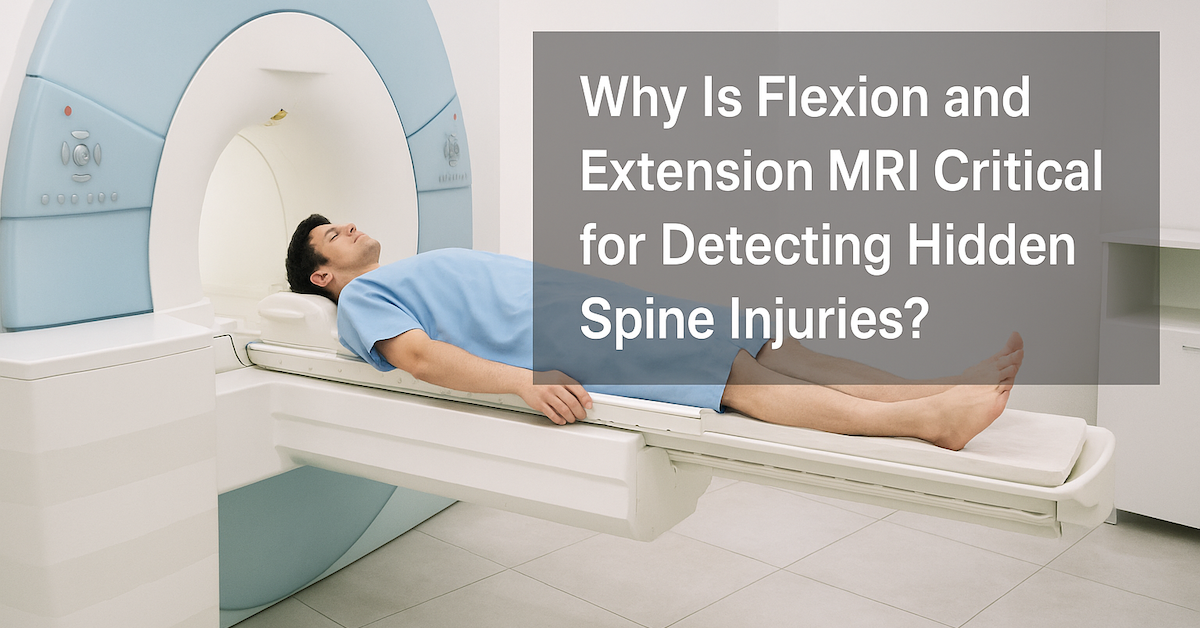
Why Is Flexion and Extension MRI Critical for Detecting Hidden Spine Injuries?
Spine injuries are often complex, and not all of them show up on traditional imaging tests. Standard MRIs provide static images of the spine while the patient lies still, which is useful for many conditions but may not capture the full picture. Some injuries only reveal themselves when the spine moves. This is where flexion and extension MRI becomes critical. By imaging the spine in different positions, this specialized scan can uncover hidden problems that a regular MRI might miss. In this article, we’ll explain what flexion and extension MRI is, why it’s so valuable, and who may benefit from it.
What Is a Flexion and Extension MRI?
A flexion and extension MRI is a diagnostic imaging test that examines the spine while the patient bends forward (flexion) and leans backward (extension). Unlike a traditional MRI, which captures images in one static position, this type of scan shows how the spine looks under movement and stress.
The goal is to reveal abnormalities that appear only when the spine is in motion. This dynamic approach provides doctors with additional details about stability, alignment, and the behavior of spinal structures that are not always visible in a standard MRI.
Why Are Some Spine Injuries Hard to Detect with Standard MRI?
A traditional MRI is excellent for identifying disc herniations, nerve compression, and other structural problems. However, because the patient lies still, the images may not show how those structures behave during movement.
Some injuries, such as spinal instability, ligament strain, or subtle disc bulges, become more obvious when the spine is in flexion or extension. Without this added perspective, patients may continue to experience pain even though their standard MRI results appear normal. This is why flexion and extension MRI is considered essential for uncovering hidden conditions.
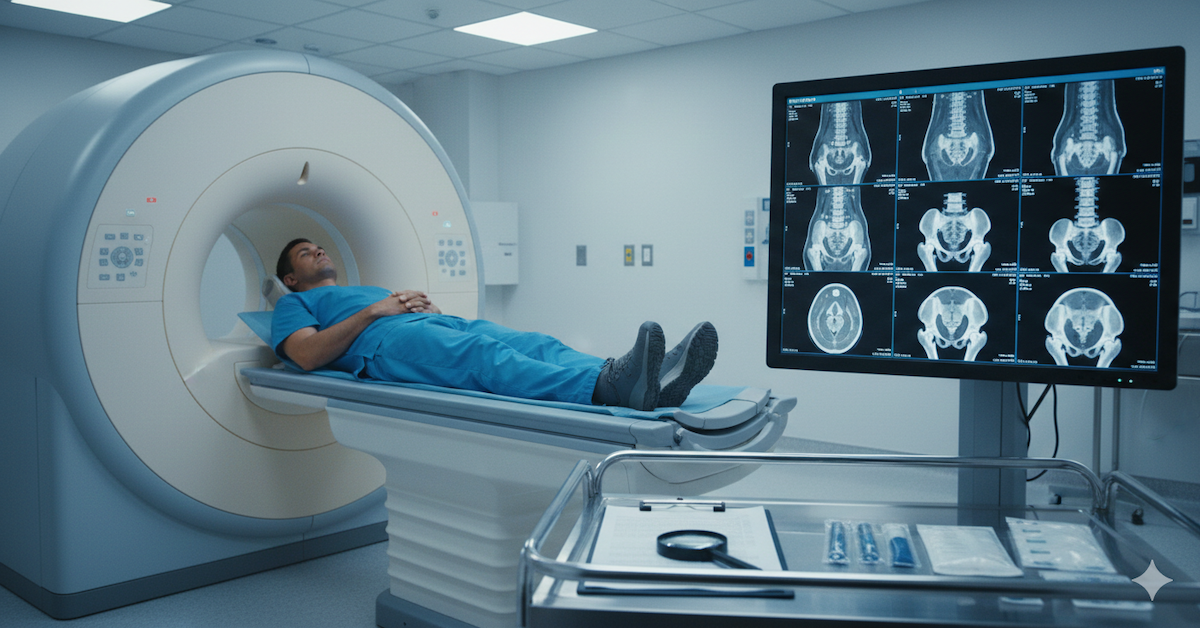
What Conditions Can Flexion and Extension MRI Reveal?
This specialized scan can detect several spine issues that static imaging might overlook:
- Spinal instability: Small shifts in vertebrae that occur only during movement.
- Ligament injuries: Strains or tears that are aggravated when bending or extending.
- Disc problems: Herniations or bulges that worsen with posture changes.
- Post-surgical complications: Instability or hardware issues that may not show up on regular scans.
By identifying these conditions early, doctors can create treatment plans that directly address the root of a patient’s symptoms.
How Does This Imaging Help Patients?
The biggest benefit of flexion and extension MRI is accuracy. When doctors have a clearer picture of what’s happening inside the spine, they can make better decisions about treatment. For some patients, this may mean avoiding unnecessary procedures because the problem is identified correctly the first time. For others, it may mean finding the hidden source of chronic pain that has gone undiagnosed.
Accurate diagnosis also leads to more targeted therapies. Whether it’s surgery, physical therapy, or pain management, knowing exactly where and how the spine is affected ensures patients receive the most effective care possible.
Who Should Consider Flexion and Extension MRI?
This type of MRI isn’t needed for everyone, but it can be especially useful for certain patients:
- Individuals with persistent back or neck pain despite normal standard MRI results.
- Patients recovering from spinal surgery who may have lingering instability.
- People with suspected ligament damage after an accident or sports injury.
- Anyone with symptoms that worsen when bending, twisting, or changing posture.
If you fall into one of these categories, your doctor may recommend a flexion and extension MRI to provide more detailed information.
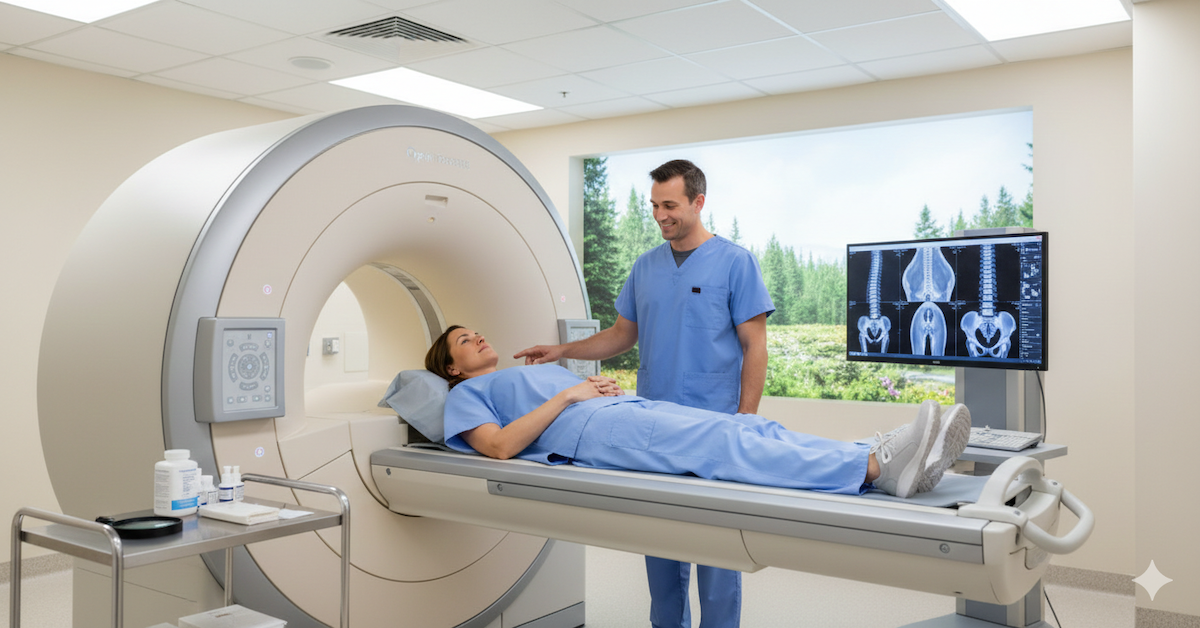
What Is the Procedure Like for Patients?
A flexion and extension MRI is similar to a standard MRI in many ways. The main difference is that the patient will be asked to move into specific positions during the scan. Typically, this involves leaning forward for flexion and leaning back for extension while images are captured.
The process is safe, non-invasive, and usually completed in less than an hour. Patients may feel some mild discomfort when holding certain positions, especially if they already have pain, but the scan itself does not cause injury. Afterward, the images are reviewed by a radiologist who provides a detailed report for the doctor.
Conclusion
Flexion and extension MRI is a critical tool for detecting hidden spine injuries that standard imaging may miss. By showing how the spine behaves under movement, it uncovers issues like instability, ligament damage, and subtle disc problems. This leads to more accurate diagnoses, better treatment plans, and greater peace of mind for patients struggling with unexplained back or neck pain.
For patients in the Chicago area, Upright MRI of Deerfield offers advanced imaging services, including flexion and extension MRI, in a comfortable and patient-focused environment. Their commitment to precision and care ensures that you get the answers you need to move forward with confidence.
SHARE THIS POST:
Leave a Comment:

The World's Most Patient-Friendly MRI. A comfortable, stress-free, and completely reliable MRI scan. We offer patients an open, upright, standup MRI experience that helps those who are claustrophobic and stress being in a confined area. Upright MRI of Deerfield is recognized as the world leader in open MRI innovation,
Our Recent Post


READ PATIENT TESTIMONIALS
Upright MRI of Deerfield.
Susan D.,
Highland Park, 39
I am going to tell everyone about your office! This was a great experience after I panicked in other MRI machines and had to leave. Thank you so much.

Judith B.,
Milwaukee, 61
I suffer from vertigo and other MRIs do not work. This was wonderful…absolutely NO discomfort at all. The MRI was so fast…I wanted to stay and watch the movie! Mumtaz was great. His humor really put me at ease. I’ve already recommended Upright MRI to friends.
Delores P.,
Glencoe, 55
Everything is so nice and professional with your place. I have been there a couple of times. My husband and I would not go anywhere else.

