
UPRIGHT MRI network BLOG

An upright brain MRI captures cerebrospinal fluid under real gravitational load. Learn how this improves CSF analysis for Chiari, CCI, and related conditions.

MRI insurance coverage is more complicated than most patients expect. Learn what affects coverage, when self-pay makes sense, and how to navigate your options.

Find out if an upright MRI reveals foraminal narrowing more accurately than a standard scan. Understand why spinal position changes what imaging can show.

Discover why a seated MRI offers a more accurate sciatica evaluation. See how upright positioning reveals nerve compression that lying-down scans often miss

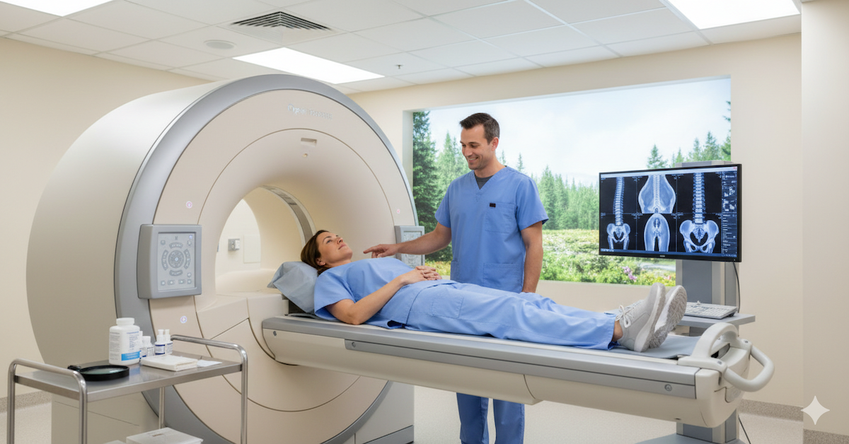
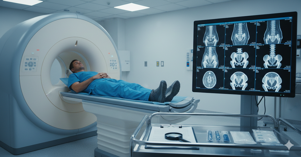
Learn what happens during an MRI scan, how long it takes, what patients feel, and how to prepare for the procedure for a smooth imaging experience.

Learn how upright MRI helps patients with anxiety and claustrophobia feel more comfortable during scans with an open design and flexible positioning.
Learn why open MRI is better for claustrophobic patients, offering more space, comfort, visibility, and a calmer experience without enclosed tubes.
Learn how a lumbar spine MRI in flexion reveals hidden issues like dynamic stenosis and nerve compression that standard lying down MRI scans may miss.
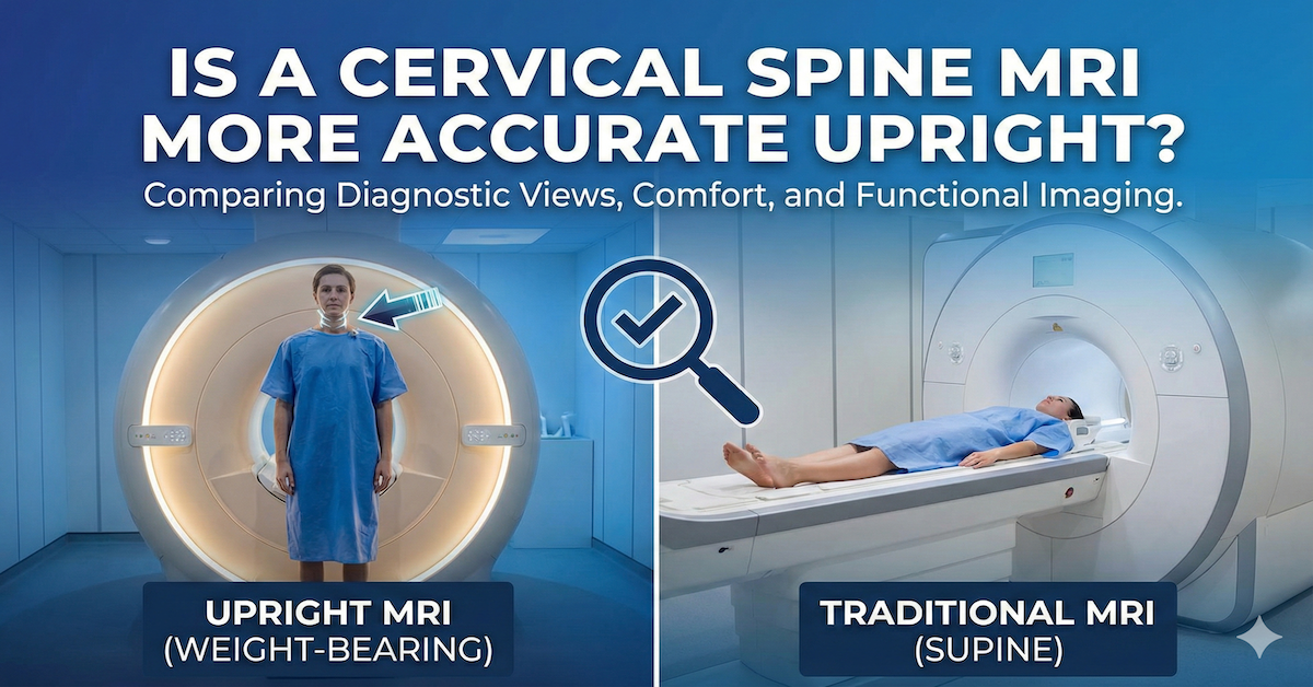
Learn why upright cervical spine MRI offers more accurate results by showing disc movement and nerve compression under natural weight and posture.

