Why Is Lateral Rotation Imaging Important for Diagnosing Spinal Conditions?
When it comes to diagnosing spine issues, getting the full picture isn't just helpful — it’s necessary. Most imaging methods, like standard MRIs or X-rays, are done while lying flat and staying perfectly still. But that’s not how we move through the world. Our spines twist, bend, carry weight, and deal with gravity. That’s where lateral rotation imaging steps in.
Lateral rotation imaging shows what’s going on in your spine while your body moves or turns. It helps doctors see what traditional scans might miss, especially when your symptoms show up only during movement.
What Is Lateral Rotation Imaging?
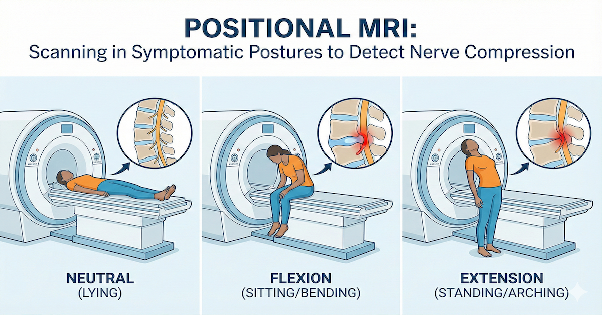
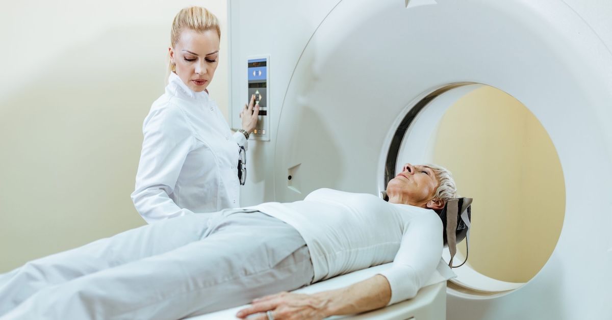
This type of imaging allows scans of the spine while the patient rotates or turns their upper body. It captures how the vertebrae, discs, and spinal cord behave during that motion. Instead of freezing your body into one position, lateral rotation scans take a dynamic view.
This is especially useful for conditions that don’t show up clearly when you’re lying still, like certain types of disc bulges, facet joint problems, or nerve impingement that only occur when you twist or lean.
Why Traditional MRIs Sometimes Fall Short
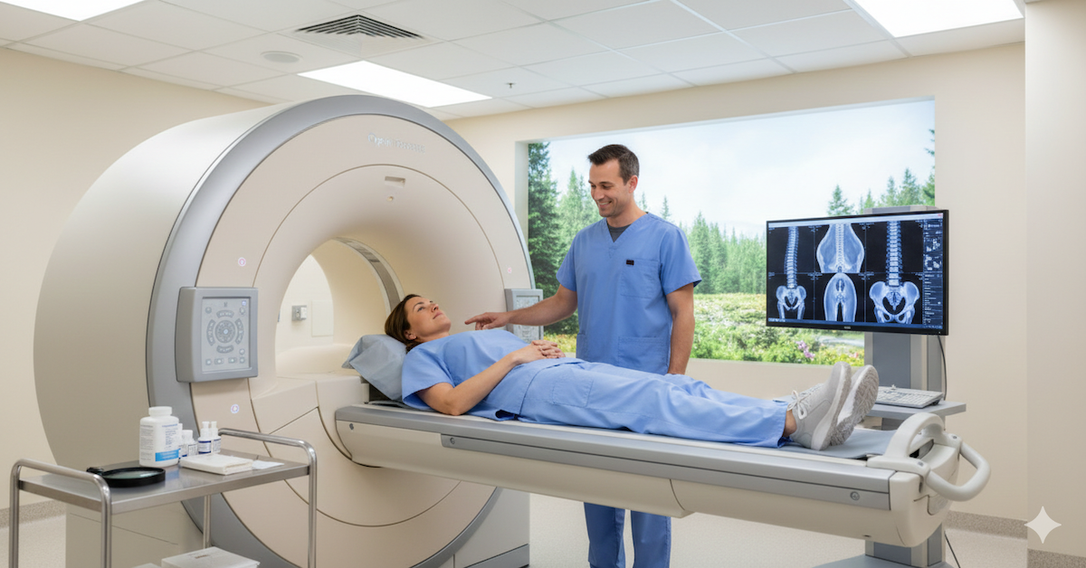
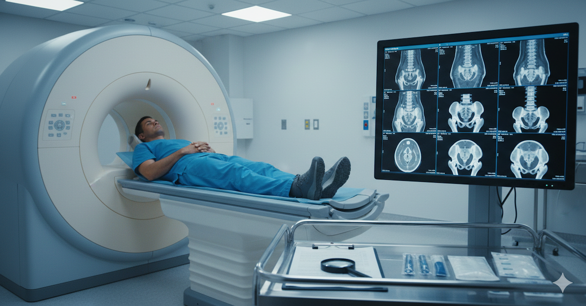
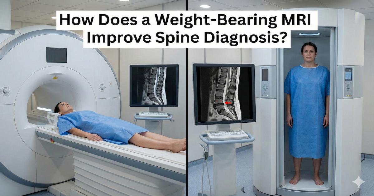
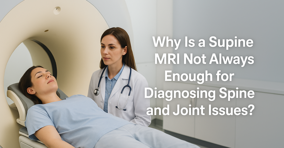
Traditional MRIs are excellent at capturing detailed images of soft tissues, discs, and the spinal cord. But the catch is that you're lying flat and motionless. That can hide problems caused by everyday movements. Pain from standing, walking, or turning might not appear during a standard MRI.
In cases like this, a traditional MRI might come back looking “normal” even though the patient is in pain. Without movement, certain disc herniations might retract slightly or spinal alignment might appear fine. So the actual cause of discomfort can go undetected unless movement is part of the scan.
Real-World Benefits of Lateral Rotation Imaging
Imagine someone has chronic low back pain that only kicks in when they turn to one side. A basic MRI shows no issues. But with lateral rotation imaging, it becomes clear that a disc bulges out or a nerve is pinched when they twist. That’s powerful diagnostic insight that can change the treatment plan completely.
Here are some common conditions where lateral rotation imaging makes a real difference:
- Spinal instability: Movement between vertebrae that shouldn’t happen
- Facet joint dysfunction: Small joints in the spine that can cause pain when twisted
- Disc displacement or herniation: Especially if only triggered by motion
- Post-surgical assessments: To check if spinal hardware holds during movement
Better Information, Better Treatment Plans
When a doctor sees exactly how your spine moves, they can recommend better treatments. For example, physical therapy might be customized based on how certain movements affect your spine. Or in more serious cases, surgery might be targeted more precisely based on actual function, not just static images.
It can also help avoid unnecessary procedures. If a patient’s pain isn't caused by structural instability, surgery might be avoided entirely in favor of a more conservative treatment like chiropractic care or pain management.
Who Should Consider Lateral Rotation Imaging?
This kind of imaging is especially useful if:
- You experience pain or discomfort during certain motions
- Your traditional MRI didn’t find the problem
- You’ve had spinal surgery and need a functional follow-up scan
- Your doctor suspects spinal instability or dynamic issues
- You need a clearer diagnosis before starting a new treatment
How Upright MRI Makes This Possible
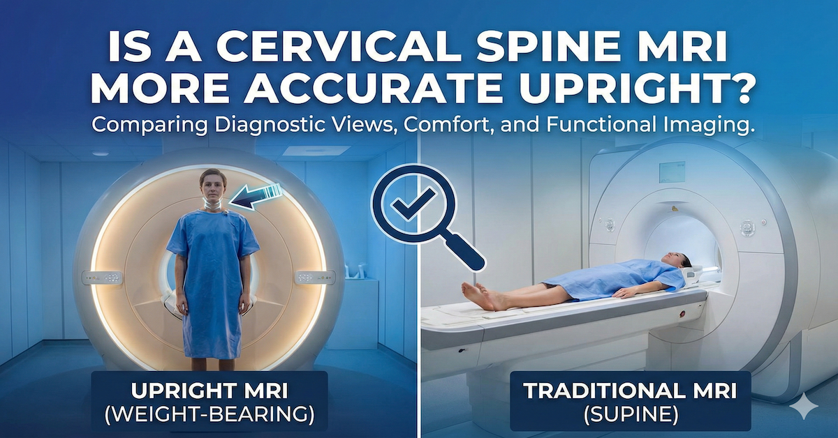
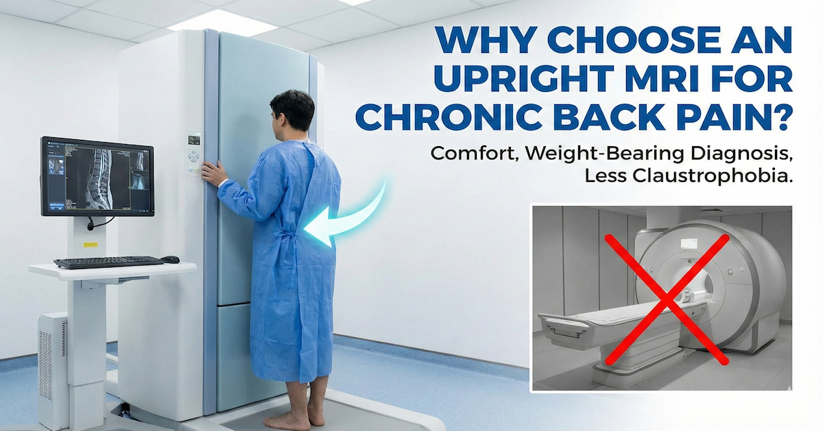
Lateral rotation imaging isn’t something every MRI machine can do. Most standard machines require you to lie flat, stay still, and remain inside a closed tube. But upright MRI allows for real-time movement while the scan takes place. That’s the key to capturing these rotating images.
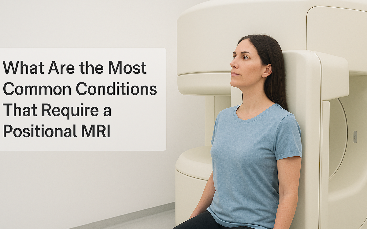
Upright MRIs let patients sit or stand and gently rotate while the scanner captures every detail. That open setup not only allows motion but also helps patients feel more relaxed during the scan.
Final Thoughts
Your spine isn’t static — it twists, turns, and reacts to the world around you. So why settle for scans that don’t show how it actually works? Lateral rotation imaging gives doctors the tools they need to see what’s really happening in motion, offering better diagnoses and smarter treatment options.
At Upright MRI of Deerfield, we offer specialized upright imaging that includes lateral rotation views for a more complete understanding of spinal conditions. Our team is here to help you uncover the real cause of your discomfort — not just how it looks lying down, but how it acts when you're living your life. If you’ve been struggling with spine issues and still don’t have answers, we’re ready to help.
SHARE THIS POST:
Leave a Comment:

The World's Most Patient-Friendly MRI. A comfortable, stress-free, and completely reliable MRI scan. We offer patients an open, upright, standup MRI experience that helps those who are claustrophobic and stress being in a confined area. Upright MRI of Deerfield is recognized as the world leader in open MRI innovation,
Our Recent Post
READ PATIENT TESTIMONIALS
Upright MRI of Deerfield.
Susan D.,
Highland Park, 39
I am going to tell everyone about your office! This was a great experience after I panicked in other MRI machines and had to leave. Thank you so much.

Judith B.,
Milwaukee, 61
I suffer from vertigo and other MRIs do not work. This was wonderful…absolutely NO discomfort at all. The MRI was so fast…I wanted to stay and watch the movie! Mumtaz was great. His humor really put me at ease. I’ve already recommended Upright MRI to friends.

Delores P.,
Glencoe, 55
Everything is so nice and professional with your place. I have been there a couple of times. My husband and I would not go anywhere else.


