
Can an Upright MRI Help Identify Shoulder Pathologies More Accurately?
Shoulder pain can disrupt everyday activities, from lifting groceries to reaching overhead. For many people, figuring out the cause of that pain is the first step toward proper treatment. While traditional MRI scans have been a standard tool for diagnosing shoulder injuries, they are not always perfect. Upright MRI technology offers a different approach, one that can sometimes reveal problems traditional methods might miss. This article explores how upright MRI works, what makes it unique, and why it may provide clearer answers for shoulder conditions.

What Is an Upright MRI?
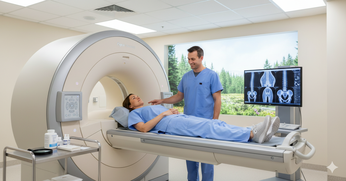
An upright MRI is a type of magnetic resonance imaging that allows patients to be scanned while sitting, standing, or in other natural positions. Instead of lying flat in a closed tube, the patient can be positioned in ways that mimic everyday movement. This feature is especially useful for joints like the shoulder, where certain issues may only appear when the joint is under normal weight-bearing conditions.
How Does Upright MRI Work for Shoulder Imaging?
When scanning the shoulder, upright MRI lets the technologist position the patient’s arm in various ways to get a complete view of the joint. This can include placing the arm overhead, by the side, or in a rotated position. By capturing images in these functional postures, the scan can highlight structural changes, instability, or impingements that might not be visible when lying down.
Common Shoulder Pathologies and Why They Can Be Hard to Diagnose
The shoulder is a complex joint made up of bones, muscles, ligaments, and tendons. Common problems include rotator cuff tears, labral tears, arthritis, bursitis, and instability. Traditional MRI scans can identify many of these issues, but some conditions change or worsen with movement or when the joint is loaded by gravity. For example, a partial tear or impingement may look minor when the patient is lying down, but upright imaging can reveal how it affects joint function in real life.
Why Traditional MRI May Miss Certain Issues
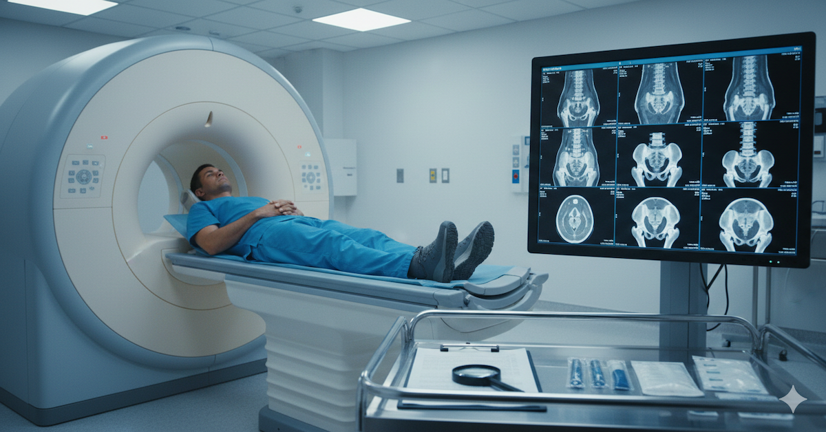
When you lie flat for a traditional MRI, the shoulder is in a relaxed, non-weight-bearing position. This can reduce the visibility of some pathologies, particularly those caused by compression or instability that occurs during regular activities. Without the effects of gravity or movement, certain soft tissue problems might appear less severe or even be overlooked entirely. Upright MRI can address this gap by scanning the joint under conditions closer to everyday use.
Advantages of Upright MRI for Shoulder Pathologies
One of the main benefits of upright MRI is its ability to show the shoulder in action. By scanning in multiple positions, radiologists can see how bones, tendons, and ligaments interact during movement. This approach can provide more accurate detection of rotator cuff tears, labral damage, and other soft tissue injuries. It also helps reveal impingements, where tendons or soft tissues are pinched during certain motions.
For patients preparing for surgery, upright MRI can be valuable for pre-surgical planning. Surgeons can get a better map of the injury, understand how it behaves in different positions, and choose the most effective repair method.
Is Upright MRI More Comfortable for Shoulder Injury Patients?
Comfort can be a challenge for patients with shoulder pain. Lying flat for a traditional MRI may cause discomfort or even increase pain, making it harder to stay still for the duration of the scan. Upright MRI often allows for a more relaxed experience, with open space and less confinement. This can be helpful for people with claustrophobia or those who cannot tolerate lying down for long periods.
How Long Does an Upright MRI Take Compared to Traditional MRI?
In most cases, an upright MRI scan takes about the same amount of time as a traditional MRI. The difference lies in the positioning process, as patients may be placed in multiple poses to capture all necessary images. The result is a more complete assessment, even if it requires a few extra minutes in the scanner.
Conclusion
An upright MRI can be a powerful tool for diagnosing shoulder pathologies more accurately. By capturing images in functional, weight-bearing positions, it can reveal issues that a standard lying-down MRI might miss. This approach can lead to better diagnosis, more effective treatment planning, and improved patient outcomes.
At Upright MRI of Deerfield, we provide advanced upright MRI imaging with a focus on patient comfort and accurate results. Our technology and experienced team ensure your shoulder is evaluated in the most natural positions possible, giving your physician the clearest view for diagnosis and treatment planning. If you are dealing with ongoing shoulder pain or need a detailed assessment before surgery, our upright MRI service can help give you the answers you need.
SHARE THIS POST:
Leave a Comment:

The World's Most Patient-Friendly MRI. A comfortable, stress-free, and completely reliable MRI scan. We offer patients an open, upright, standup MRI experience that helps those who are claustrophobic and stress being in a confined area. Upright MRI of Deerfield is recognized as the world leader in open MRI innovation,
Our Recent Post


READ PATIENT TESTIMONIALS
Upright MRI of Deerfield.
Susan D.,
Highland Park, 39
I am going to tell everyone about your office! This was a great experience after I panicked in other MRI machines and had to leave. Thank you so much.

Judith B.,
Milwaukee, 61
I suffer from vertigo and other MRIs do not work. This was wonderful…absolutely NO discomfort at all. The MRI was so fast…I wanted to stay and watch the movie! Mumtaz was great. His humor really put me at ease. I’ve already recommended Upright MRI to friends.
Delores P.,
Glencoe, 55
Everything is so nice and professional with your place. I have been there a couple of times. My husband and I would not go anywhere else.

