What Is Covered in an MRI for Foot Conditions?
MRI technology has become a game-changer in diagnosing various foot conditions. This non-invasive imaging technique allows doctors to get a detailed view of the structures within your foot, helping to pinpoint the cause of pain or dysfunction. If you've been scheduled for an MRI to investigate foot problems, here's a comprehensive guide to help you understand what to expect and what the MRI covers.
Basics of MRI Technology
MRI, or Magnetic Resonance Imaging, uses powerful magnets and radio waves to create detailed images of the inside of your body. Unlike X-rays and CT scans, MRI does not use ionizing radiation, making it a safer option for many patients. This imaging technique is particularly useful for soft tissues, which are often involved in foot conditions.

Common Foot Conditions Diagnosed with MRI
An MRI can reveal a wide range of foot conditions, thanks to its ability to provide detailed images of both bones and soft tissues. Some common conditions that an MRI can help diagnose include:
- Plantar Fasciitis: Inflammation of the plantar fascia, the thick band of tissue that runs across the bottom of your foot.
- Achilles Tendinitis: Inflammation of the Achilles tendon, which connects the calf muscles to the heel bone.
- Stress Fractures: Tiny cracks in the bones that can develop from overuse or repetitive activity.
- Ligament Sprains and Tears: Injuries to the ligaments, which are the tough bands of tissue that connect bones together.
- Arthritis: Inflammation of the joints, which can cause pain and stiffness.
- Morton’s Neuroma: A thickening of the tissue around one of the nerves leading to your toes.
- Tarsal Tunnel Syndrome: Compression of the tibial nerve as it travels through the tarsal tunnel, leading to pain and tingling in the foot.
Preparing for an MRI
Knowing what to expect can help ease any anxiety you might have about getting an MRI. Here's a brief overview of the process:
- Preparation: You might need to change into a hospital gown and remove any metal objects, as metal can interfere with the magnetic field.
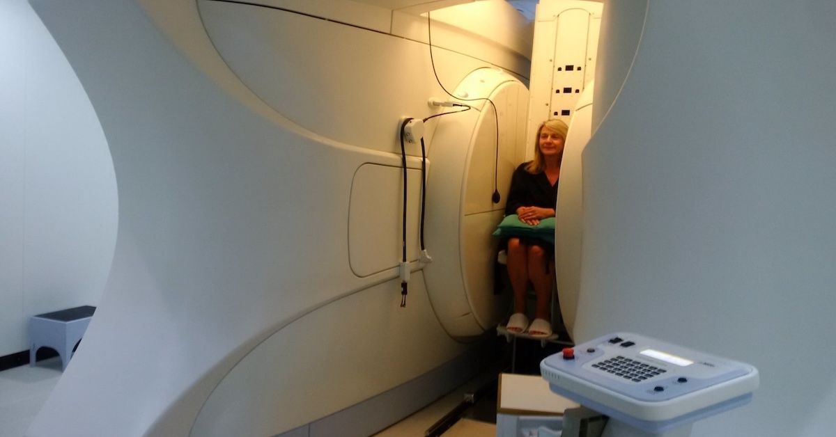
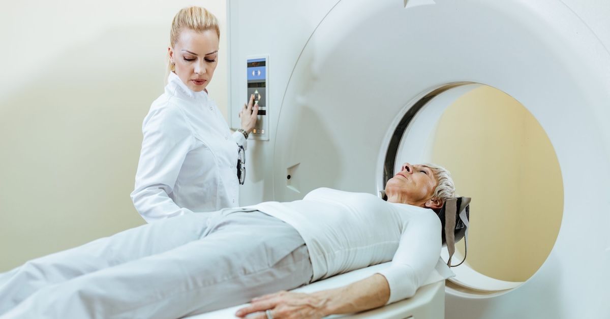
- Positioning: For a foot MRI, you will likely lie down on the MRI table with your foot positioned in the scanner. Comfort is key, as you'll need to remain still during the scan.
- Duration: The scan typically takes 30-60 minutes. To make the experience more comfortable, you might be offered earplugs or headphones, as the machine can be noisy.
MRI Imaging Techniques for Foot Conditions
MRI scans use different sequences to highlight various structures within the foot:
- T1 and T2-weighted images: These are standard MRI sequences that provide detailed images of the foot’s anatomy.
- STIR (Short Tau Inversion Recovery) sequences: Particularly useful for detecting inflammation and edema.
- Contrast-enhanced MRI: Sometimes, a contrast agent is injected to enhance the visibility of certain tissues or abnormalities.
Detailed Structures Examined in a Foot MRI
An MRI scan of the foot can provide detailed images of several structures:
- Bones and Joints: The MRI can visualize all the bones in the foot, including the metatarsals, phalanges, and tarsal bones (like the talus, calcaneus, navicular, cuboid, and cuneiforms). It also shows joint spaces and cartilage, which can be crucial for diagnosing arthritis or other joint conditions.
- Soft Tissues: The MRI can capture images of muscles and tendons, such as the Achilles tendon and peroneal tendons, as well as ligaments like the plantar fascia and deltoid ligament. It also reveals nerves (like the tibial and plantar nerves), blood vessels, and connective tissues.
Interpreting MRI Results for Foot Conditions
Interpreting MRI results involves understanding both normal and abnormal findings:
- Normal vs. Abnormal Findings: A healthy MRI will show well-defined structures with no signs of inflammation or abnormal fluid accumulation. Abnormal findings might include:
- Signs of Inflammation or Injury: These could show up as increased signal intensity on certain MRI sequences, indicating swelling or damage.
- Bone Marrow Edema: This appears as a bright area within the bone and often indicates stress fractures or other bone injuries.
- Tendon or Ligament Tears: Partial or complete tears will be visible, with disrupted or abnormal tissue patterns.
- Joint Degeneration and Arthritis: This can be seen as narrowed joint spaces, bone spurs, or cartilage loss.
Clinical Correlation and Next Steps
After receiving your MRI results, it’s important to discuss them with your healthcare provider. They will correlate the MRI findings with your symptoms and physical examination results. This holistic approach ensures that the treatment plan addresses the root cause of your foot problems. Depending on the diagnosis, potential next steps may include:
- Conservative Management: Rest, physical therapy, and changes in footwear or activity levels can often help.
- Medications: Anti-inflammatory drugs or pain relievers may be prescribed.
- Interventional Procedures: Injections, such as corticosteroids, might be recommended.
- Surgical Options: In severe cases, surgery might be necessary to correct the underlying problem.
Advantages of MRI for Foot Conditions
The benefits of using MRI to diagnose foot conditions are numerous:
- Non-invasive: No need for incisions or exposure to radiation.
- High-resolution Images: Detailed pictures of both bone and soft tissue structures.
- Comprehensive: Ability to detect a wide range of abnormalities, from inflammation to bone fractures.
Conclusion
An MRI for foot conditions provides a comprehensive view of the various structures in the foot, helping to diagnose a wide range of issues accurately. Understanding what an MRI covers and how to interpret the results can empower you to make informed decisions about your health. For expert MRI services and detailed imaging, consider Upright MRI of Deerfield. Our state-of-the-art technology and experienced team are dedicated to providing the best care and clarity for your medical needs.
SHARE THIS POST:
Leave a Comment:

The World's Most Patient-Friendly MRI. A comfortable, stress-free, and completely reliable MRI scan. We offer patients an open, upright, standup MRI experience that helps those who are claustrophobic and stress being in a confined area. Upright MRI of Deerfield is recognized as the world leader in open MRI innovation,
Our Recent Post
READ PATIENT TESTIMONIALS
Upright MRI of Deerfield.
Susan D.,
Highland Park, 39
I am going to tell everyone about your office! This was a great experience after I panicked in other MRI machines and had to leave. Thank you so much.

Judith B.,
Milwaukee, 61
I suffer from vertigo and other MRIs do not work. This was wonderful…absolutely NO discomfort at all. The MRI was so fast…I wanted to stay and watch the movie! Mumtaz was great. His humor really put me at ease. I’ve already recommended Upright MRI to friends.

Delores P.,
Glencoe, 55
Everything is so nice and professional with your place. I have been there a couple of times. My husband and I would not go anywhere else.

