
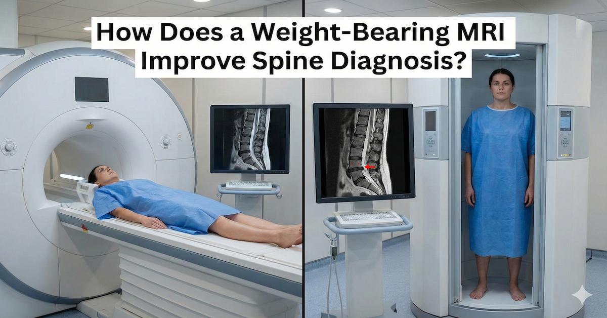
How Does a Weight Bearing MRI Improve Spine Diagnosis?
Spine pain can feel confusing when symptoms appear only while standing, sitting, or moving. Many people go through traditional MRI scans and still do not get clear answers because the images are taken while lying flat. This position often removes the pressure that causes real symptoms. A weight bearing MRI gives a more natural view of your spine by capturing images while your body is upright or partially upright.
This type of scan helps doctors understand how gravity affects your discs, joints, nerves, and alignment. When the spine carries weight, it behaves very differently than it does when you lie down. A weight bearing scan reveals these changes in a way that a standard MRI cannot.
Quick answer: A weight bearing MRI improves spine diagnosis by showing how the spine looks and functions under real life pressure, helping doctors detect disc issues, nerve compression, misalignment, and posture related problems that lying down MRI scans can miss.
Understanding What a Weight Bearing MRI Is
A weight bearing MRI is an imaging method that allows the patient to be scanned while sitting, standing, or positioned upright. Instead of lying on a flat table, the body stays in a natural position with gravity applied. This gives a clearer view of how the spine actually functions in daily life.
The machine is designed so patients can sit or stand comfortably while the scanner rotates around them. This upright position offers a more functional image of the spine and often reveals problems that do not show up when the body is completely relaxed.
How a Weight Bearing MRI Works
The scanner captures detailed images of the spine while it is supporting weight. Because the spine compresses and shifts under pressure, the images show how discs move, how vertebrae align, and whether nerves are being pinched during normal activity. This creates a more realistic picture of what causes pain or discomfort.
Why Upright Imaging Matters for Spinal Conditions
Many spinal problems do not appear when the pressure is removed from the body. Herniated discs often flatten when lying down. Misaligned vertebrae may look normal when gravity is not applied. Even nerve compression can lessen when the spine is horizontal. Weight bearing MRI brings these issues to the surface so they can be diagnosed correctly.
Key Differences Between a Weight Bearing MRI and a Traditional MRI
The main difference is positioning. A traditional MRI requires the patient to lie flat, which relaxes the spine and removes the very pressure that may be causing symptoms. This can lead to scans that show normal results even when the patient still feels pain.
A weight bearing MRI keeps the spine in its natural working position. It captures functional images that show how the spine behaves during sitting, standing, bending, or leaning. Because posture affects spinal alignment, these images can reveal issues a standard scan cannot detect.
Functional Imaging Advantages
A weight bearing scan can be taken in multiple positions. This allows doctors to see how discs and nerves respond to bending, extending, or sitting upright. These flexible views make diagnosis more precise.
How Weight Bearing MRI Improves Spine Diagnosis
Weight bearing MRI is especially valuable for conditions that change with posture. When gravity is applied, discs compress, joint spaces narrow, and nerve openings become smaller. These changes help doctors locate the true source of pain.
More Accurate Detection of Herniated or Bulging Discs
Disc material may press on nerves only when the body is upright. A standard MRI may show nothing unusual, but a weight bearing scan can reveal the disc bulge clearly.
Identifying Spinal Instability and Misalignment
Conditions such as spondylolisthesis become easier to diagnose when the spine is under load. An upright scan shows how much the vertebrae shift when the patient is standing or sitting.
Clearer Visualization of Nerve Compression
Gravity can reduce the space where nerves exit the spine. Weight bearing imaging shows exactly when and where the nerves become compressed.
Better Evaluation of Chronic Pain Causes
People who feel pain only while walking, sitting, or standing benefit greatly from this type of scan. Weight bearing MRI identifies the pressure points that produce those symptoms.
Who Benefits Most From a Weight Bearing MRI
Patients who feel pain during standing or walking often see the most benefit. Upright imaging also supports athletes, people with posture related problems, and patients whose traditional MRI results did not match their symptoms.
Common Conditions Best Diagnosed With Weight Bearing MRI
These include sciatica, degenerative disc disease, foraminal stenosis, spondylolisthesis, facet joint issues, and posture driven alignment problems.
What to Expect During a Weight Bearing MRI
Patients are usually positioned sitting or standing. The scan is comfortable and open, which helps reduce anxiety. The scanner takes images over several minutes while you remain still. Because the design is more open than traditional MRI machines, many patients find this method easier.
Is a Weight Bearing MRI More Effective Than a Standard MRI?
In many cases, yes. If your symptoms change with posture, an upright scan provides far better information. However, for certain soft tissue injuries that are not affected by position, a standard MRI may still be enough.
Conclusion
A weight bearing MRI gives a more complete and realistic view of how your spine functions under daily pressure. It reveals disc problems, nerve compression, and alignment issues that traditional lying down scans often miss. This leads to clearer diagnosis and more effective treatment planning.
If you want accurate imaging that reflects how your spine truly behaves, UpRight MRI of Deerfield offers advanced weight bearing MRI technology that helps you get real answers and better care.
SHARE THIS POST:
Leave a Comment:

The World's Most Patient-Friendly MRI. A comfortable, stress-free, and completely reliable MRI scan. We offer patients an open, upright, standup MRI experience that helps those who are claustrophobic and stress being in a confined area. Upright MRI of Deerfield is recognized as the world leader in open MRI innovation,
Our Recent Post


READ PATIENT TESTIMONIALS
Upright MRI of Deerfield.
Susan D.,
Highland Park, 39
I am going to tell everyone about your office! This was a great experience after I panicked in other MRI machines and had to leave. Thank you so much.

Judith B.,
Milwaukee, 61
I suffer from vertigo and other MRIs do not work. This was wonderful…absolutely NO discomfort at all. The MRI was so fast…I wanted to stay and watch the movie! Mumtaz was great. His humor really put me at ease. I’ve already recommended Upright MRI to friends.
Delores P.,
Glencoe, 55
Everything is so nice and professional with your place. I have been there a couple of times. My husband and I would not go anywhere else.

