
Can a Positional MRI Detect Nerve Compression Better?
Nerve compression can cause pain, numbness, tingling, or weakness that changes depending on how you sit, stand, or move. Many people struggle with symptoms that come and go, yet traditional MRI scans fail to show a clear cause because the images are taken while lying flat. When pressure is removed from the spine, the problem often disappears from the scan.
A positional MRI solves this issue by capturing images of the spine in different positions. This makes it easier to see how posture and movement affect nerve pathways.
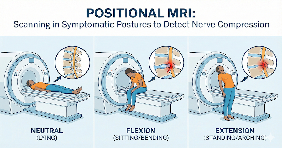
Quick answer: A positional MRI can detect nerve compression better because it shows the spine in real life positions, such as sitting, standing, bending, or rotating, revealing nerve pinching that traditional lying down MRIs often miss.
What a Positional MRI Is
A positional MRI is an imaging method that allows the patient to be scanned in several postures instead of lying down. The goal is to show how the spine behaves when gravity and movement influence its alignment.
How a Positional MRI Works
The scanner can adjust to upright, seated, forward bending, or rotated positions. As the patient shifts posture, the MRI captures detailed images of the discs, joints, and nerves. These images reflect how the spine behaves during daily movement.
Why Position Changes Reveal Hidden Spine Problems
Many spinal issues disappear when the body lies flat. Gravity no longer compresses the discs or narrows nerve pathways. Positional MRI exposes these changes by capturing the spine exactly as it functions during normal activity. This makes it easier to see when and where nerves are being pinched.
How Positional MRI Helps Detect Nerve Compression
A positional MRI is especially useful for conditions that change with posture or movement.
Showing Compression That Appears Only in Certain Positions
Some people feel pain only when standing, bending, or sitting for long periods. A positional MRI recreates those positions so doctors can observe how nerve spaces tighten or shift.
Real Time Changes in Disc Height and Joint Space
Discs and joints adjust slightly with every posture change. These shifts can increase or decrease pressure on nerves. Positional imaging captures these changes clearly.
Improved Identification of Foraminal Stenosis
The foramina are small openings where nerves exit the spine. These openings can shrink during standing, leaning, or rotation. Positional MRI shows this narrowing, which is often missed in traditional scans.
Common Symptoms Positional MRI Helps Explain
Many posture related symptoms become easier to diagnose with positional imaging, including:
• Pain during sitting or standing
• Radiating pain in the arms or legs
• Tingling or numbness triggered by movement
• Weakness or discomfort that appears only in certain positions
These symptoms often confuse patients because they appear inconsistent. Positional MRI brings clarity.
Conditions Best Evaluated With Positional MRI
Certain spinal conditions become more visible when posture changes. These include:
• Herniated discs that shift during bending
• Foraminal stenosis
• Central canal stenosis
• Spondylolisthesis and spinal instability
• Posture related nerve compression
These conditions may look mild or invisible on a traditional MRI, yet become obvious when gravity is applied.
Positional MRI vs Traditional MRI for Nerve Compression
A traditional MRI captures static images while the patient is lying down. This position removes natural pressure from the spine, which means nerve compression may no longer be visible.
A positional MRI provides dynamic imaging. It shows how spinal structures move and compress during everyday posture. For many patients, this reveals the true cause of their symptoms.
Advantages for Treatment Planning
When doctors can pinpoint the exact movement or posture that causes nerve compression, treatment becomes more precise. Positional MRI helps guide:
• Physical therapy plans
• Chiropractic care
• Injection therapy
• Minimally invasive procedures
• Surgical decision making
Better imaging leads to better outcomes.
What Patients Can Expect During a Positional MRI
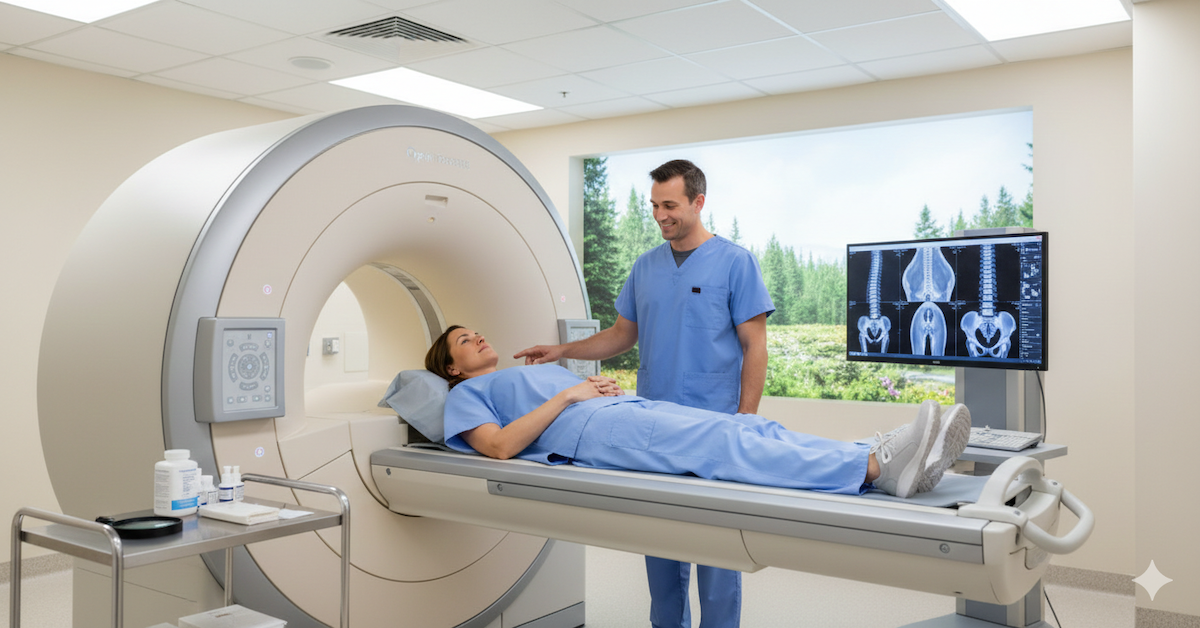
A positional MRI is more open and comfortable than a traditional MRI. Patients remain upright or partially upright, which helps reduce feelings of confinement. The scan takes a similar amount of time, but the experience often feels easier because of the open design and natural body position.
Patients will be guided into positions that match their symptoms. If pain appears when bending or sitting, the radiologist may capture images in that exact posture.
Is Positional MRI Always the Better Choice?
Positional MRI is especially helpful when symptoms depend on posture. If nerve compression occurs only during certain movements, upright imaging will provide far clearer answers than a traditional scan.
However, for soft tissue injuries or issues unrelated to gravity or movement, a standard MRI may still be sufficient. The best choice depends on the symptoms and the suspected condition.
Conclusion
A positional MRI offers a more realistic view of how the spine behaves during daily movement. By capturing images of the spine in sitting, standing, or bending positions, it reveals nerve compression that traditional lying down MRIs often cannot detect. For many patients, this leads to a faster, clearer, and more accurate diagnosis.
If you want imaging that matches how your body truly moves and functions, UpRight MRI of Deerfield provides advanced positional MRI technology that helps uncover the real source of pain and nerve compression.
SHARE THIS POST:
Leave a Comment:

The World's Most Patient-Friendly MRI. A comfortable, stress-free, and completely reliable MRI scan. We offer patients an open, upright, standup MRI experience that helps those who are claustrophobic and stress being in a confined area. Upright MRI of Deerfield is recognized as the world leader in open MRI innovation,
Our Recent Post


READ PATIENT TESTIMONIALS
Upright MRI of Deerfield.
Susan D.,
Highland Park, 39
I am going to tell everyone about your office! This was a great experience after I panicked in other MRI machines and had to leave. Thank you so much.

Judith B.,
Milwaukee, 61
I suffer from vertigo and other MRIs do not work. This was wonderful…absolutely NO discomfort at all. The MRI was so fast…I wanted to stay and watch the movie! Mumtaz was great. His humor really put me at ease. I’ve already recommended Upright MRI to friends.
Delores P.,
Glencoe, 55
Everything is so nice and professional with your place. I have been there a couple of times. My husband and I would not go anywhere else.

