
What is the Purpose and Procedure of MRI Brain Scans?
MRI brain scans are an essential tool in modern medicine, offering detailed insights into the inner workings of the human brain. These scans serve a dual purpose: they allow healthcare professionals to visualize the anatomy of the brain with remarkable clarity, while also providing valuable information about brain function and potential abnormalities. In this article, we will explore the purpose and procedure of MRI brain scans, shedding light on why they are conducted and what patients can expect during the process.
Understanding the Purpose of MRI Brain Scans
At the heart of every MRI brain scan lies a simple yet profound goal: to help doctors see inside the brain without the need for invasive procedures. By harnessing the power of magnetic fields and radio waves, MRI technology generates highly detailed images of the brain's internal structures. These images can reveal important information about the size, shape, and condition of various brain regions, allowing doctors to diagnose a wide range of neurological conditions.
One of the primary purposes of MRI brain scans is to detect abnormalities such as tumors, lesions, or areas of abnormal tissue growth. Unlike traditional imaging techniques like X-rays or CT scans, which provide only a limited view of the brain, MRI offers unparalleled clarity and precision. This makes it an invaluable tool for identifying potentially life-threatening conditions and guiding appropriate treatment strategies.
But MRI brain scans aren't just about spotting problems they're also about understanding how the brain functions. By measuring factors like blood flow, oxygen levels, and neural activity, MRI can provide insights into brain health and performance. This information is particularly useful in cases of neurological disorders such as stroke, Alzheimer's disease, or epilepsy, where changes in brain function may precede visible structural changes.
Exploring the Procedure of MRI Brain Scans
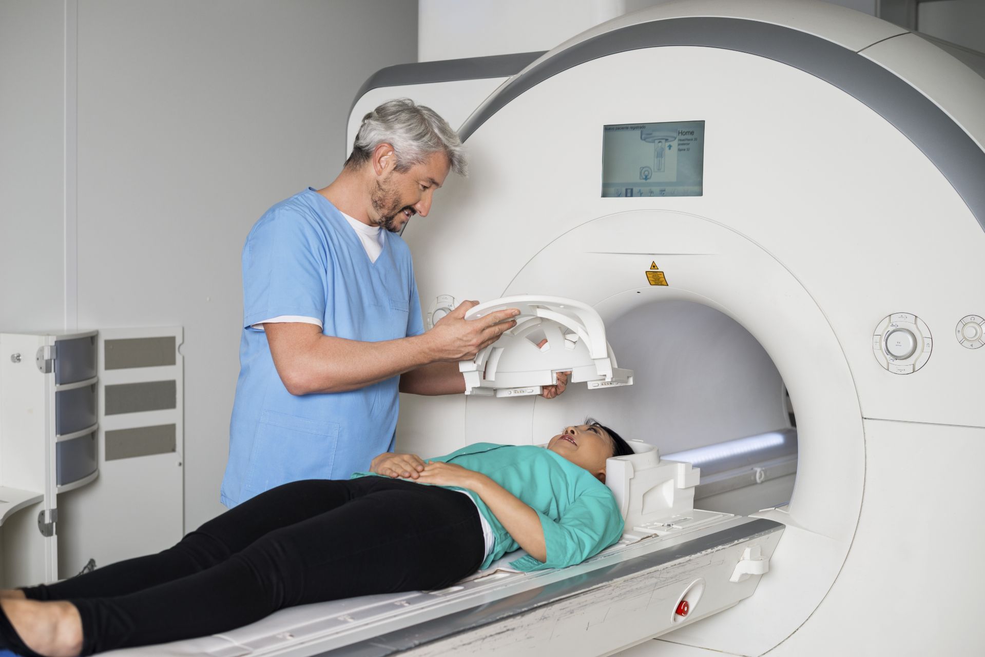
So, what exactly happens during an MRI brain scan? The procedure typically begins with some preparation, as patients are asked to remove any metal objects or jewelry that could interfere with the magnetic field. Once inside the MRI machine, patients are positioned carefully to ensure that the area of interest the brain is in the optimal position for imaging.
During the scan itself, patients may hear loud knocking or tapping noises as the MRI machine generates the images. While this can be somewhat disconcerting, it's perfectly normal and nothing to worry about. Some patients may also receive a contrast agent, a special dye injected into the bloodstream to enhance the visibility of certain structures or abnormalities.
Throughout the procedure, patient safety is of utmost importance. MRI machines produce powerful magnetic fields, so it's crucial to follow safety protocols to avoid injury or discomfort. Patients with certain medical implants or conditions may need to undergo additional screening or precautions to ensure their safety during the scan.
The Benefits and Limitations of MRI Brain Scans
The benefits of MRI brain scans are clear: they offer a safe, non-invasive way to examine the brain in exquisite detail, providing valuable information for diagnosis and treatment. Compared to other imaging techniques, MRI is particularly well-suited for studying soft tissues like the brain, offering superior contrast and resolution.
However, MRI brain scans also have their limitations. For one, they can be expensive and may not be readily available to all patients. Additionally, certain individuals may experience claustrophobia or anxiety inside the MRI machine, which can make the procedure challenging or uncomfortable.
Despite these limitations, MRI brain scans remain an indispensable tool in the field of neuroimaging, offering unparalleled insights into the structure and function of the human brain. Whether used to diagnose a medical condition, monitor disease progression, or guide surgical interventions, MRI brain scans play a vital role in modern healthcare.
In conclusion, MRI brain scans are a powerful diagnostic tool that provides invaluable information about the structure and function of the brain. By harnessing the principles of magnetic resonance imaging, doctors can visualize the brain in remarkable detail, aiding in the diagnosis and treatment of a wide range of neurological conditions. If you have any questions or concerns about MRI brain scans, don't hesitate to reach out to your healthcare provider. And remember, if you're in need of an MRI in the Deerfield area, consider Upright MRI of Deerfield for compassionate care and state-of-the-art imaging services.
SHARE THIS POST:
Leave a Comment:

The World's Most Patient-Friendly MRI. A comfortable, stress-free, and completely reliable MRI scan. We offer patients an open, upright, standup MRI experience that helps those who are claustrophobic and stress being in a confined area. Upright MRI of Deerfield is recognized as the world leader in open MRI innovation,
Our Recent Post


READ PATIENT TESTIMONIALS
Upright MRI of Deerfield.
Susan D.,
Highland Park, 39
I am going to tell everyone about your office! This was a great experience after I panicked in other MRI machines and had to leave. Thank you so much.

Judith B.,
Milwaukee, 61
I suffer from vertigo and other MRIs do not work. This was wonderful…absolutely NO discomfort at all. The MRI was so fast…I wanted to stay and watch the movie! Mumtaz was great. His humor really put me at ease. I’ve already recommended Upright MRI to friends.
Delores P.,
Glencoe, 55
Everything is so nice and professional with your place. I have been there a couple of times. My husband and I would not go anywhere else.

