
Can an Upright MRI Improve the Detection of Whiplash-Related Injuries?
Whiplash injuries, commonly caused by car accidents, can lead to long-term pain and discomfort if not diagnosed correctly. While whiplash might seem like a straightforward neck injury, the symptoms can vary, and the damage isn’t always visible on traditional diagnostic imaging. That’s where Upright MRI technology comes in. For years, conventional MRIs have been the gold standard in diagnosing injuries, but they have limitations when it comes to detecting injuries like whiplash. By allowing patients to be scanned in an upright position, Upright MRIs provide a more accurate view of the injury’s impact, offering a significant advantage for those dealing with whiplash-related conditions.
What Is Whiplash and Why Is It Hard to Diagnose?
Whiplash occurs when the neck is suddenly jolted forward and backward, often as a result of a rear-end car collision. This jerking motion can strain the muscles, ligaments, and tendons in the neck, leading to symptoms like pain, stiffness, headaches, dizziness, and even shoulder pain. The severity of whiplash can range from mild to severe, and in many cases, the soft tissue damage isn’t immediately visible on X-rays or traditional MRI scans. This makes diagnosing whiplash tricky since its effects often don’t show up on imaging tests designed for bone injuries.
Traditional MRI machines can’t replicate the motion or weight-bearing conditions the neck experiences during whiplash. This limitation means that subtle soft tissue injuries like sprained ligaments or damaged discs might be overlooked, leaving the patient without a clear diagnosis and the right treatment plan.
Traditional MRI vs. Upright MRI for Whiplash Detection
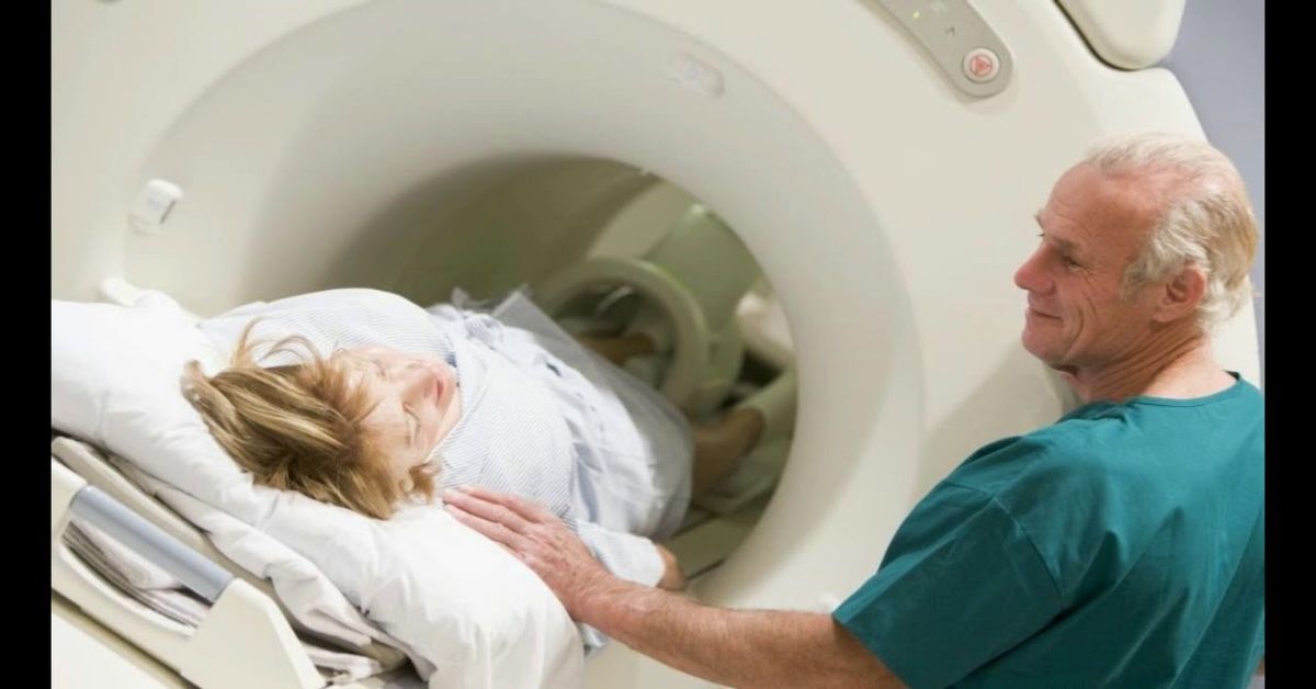
Traditional MRIs are conducted with the patient lying flat on their back, which doesn’t mimic the movement or stress the neck experiences during whiplash. While traditional MRI machines offer great detail in static images, they miss the dynamic aspect of an injury that occurs during movement. This can make it difficult to capture the full extent of whiplash-related damage, especially when ligaments or muscles are involved.
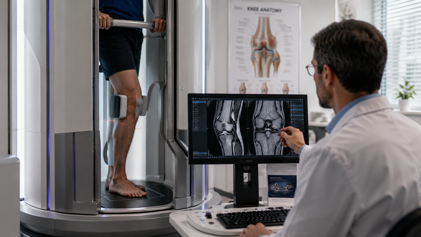
Upright MRI offers a more comprehensive approach. With this technology, the patient is positioned upright, in a natural, weight-bearing posture. This position allows the neck to be subjected to the same stresses and movements that occur during a whiplash event, offering a more accurate representation of how the neck is functioning under pressure. By capturing images in this position, Upright MRI can better detect issues like spinal misalignments, ligament sprains, or disc bulges—conditions that are common with whiplash injuries but might be missed in a traditional MRI.
Why Upright MRI Is Better for Detecting Whiplash Injuries
One of the main advantages of Upright MRI is its ability to capture the neck's movement during the scan. Traditional MRI is static, showing only a snapshot of the neck in a non-functional, resting position. For whiplash victims, this is problematic because the injury often involves the neck being subjected to fast, forceful motion. When the neck is in motion, it can put stress on soft tissues like ligaments and discs, and these stresses often go undetected in a typical MRI scan.
Upright MRI allows healthcare providers to see the effects of movement and weight-bearing forces on the neck. This makes it easier to identify damage that is otherwise invisible when the patient is lying down. For example, a herniated disc or ligament strain may not show up when the neck is in a resting position but could be clearly visible during the dynamic scanning of an Upright MRI.
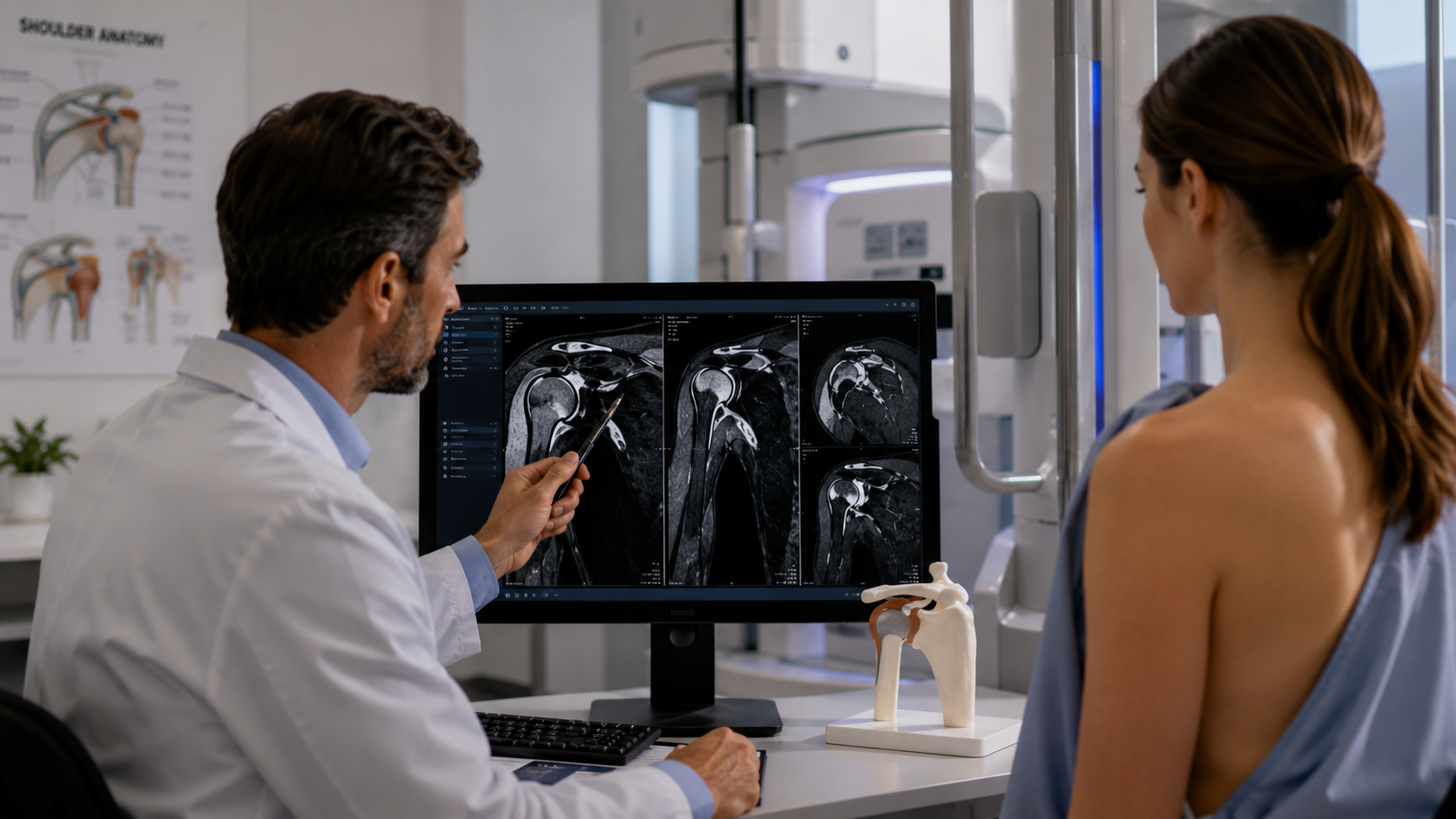
This technology can also provide more detailed imaging of the cervical spine and surrounding structures. It can show how the spine moves, helping doctors pinpoint the areas of injury more accurately and providing a better understanding of the injury’s impact on the patient’s daily life. In essence, an Upright MRI provides a much more realistic and relevant image of the injury compared to traditional methods.
Additional Benefits of Upright MRI for Whiplash Diagnosis
Upright MRI provides several advantages beyond just improving the detection of whiplash-related injuries. First, it offers a more comfortable experience for patients. Traditional MRIs often require patients to lie still in a narrow tube for an extended period, which can be uncomfortable—especially for individuals suffering from neck pain. In contrast, the Upright MRI allows patients to remain in a position that feels more natural and less constrained.
Moreover, the use of Upright MRI reduces the need for multiple tests. With traditional MRIs, doctors may have to request follow-up imaging or additional diagnostic tests to get a clearer picture of the injury. With Upright MRI, much of the necessary information can be gathered in a single session, saving time and reducing the need for additional appointments.
Another benefit of Upright MRI is its ability to help identify other neck-related problems that could be contributing to the symptoms. For example, issues like spinal stenosis, degenerative disc disease, or cervical instability can also be detected through Upright MRI, allowing for a comprehensive assessment of the neck's health.
When Is Upright MRI a Better Option?
While Upright MRI offers clear advantages in diagnosing whiplash, it’s not always necessary for every patient. For minor injuries or routine conditions, traditional MRI or X-rays may still be sufficient. However, for individuals who are suffering from chronic pain or whose symptoms don’t align with what is visible on traditional imaging, an Upright MRI may provide a more accurate diagnosis.
This technology is particularly useful for patients who experience ongoing neck pain, difficulty moving their head, or other symptoms after a traumatic event like a car accident. For those who have tried other forms of treatment without success, an Upright MRI might reveal hidden injuries that explain the continued discomfort.
Conclusion
Upright MRI is a game-changer when it comes to diagnosing whiplash-related injuries. Its ability to capture dynamic, real-world movement gives doctors a clearer picture of the injury and allows for more accurate, effective treatment plans. Whether you're dealing with subtle ligament strains, disc issues, or cervical misalignments, Upright MRI provides the detailed imaging necessary to make an accurate diagnosis.
If you’ve experienced whiplash and traditional imaging hasn’t provided the answers you need, consider exploring the benefits of Upright MRI. Upright MRI of Deerfield specializes in cutting-edge imaging that can help uncover the full extent of your injury and guide your treatment plan. With our advanced technology, we’re here to ensure that your diagnosis is as clear and comprehensive as possible, so you can get on the path to recovery.
SHARE THIS POST:
Leave a Comment:

The World's Most Patient-Friendly MRI. A comfortable, stress-free, and completely reliable MRI scan. We offer patients an open, upright, standup MRI experience that helps those who are claustrophobic and stress being in a confined area. Upright MRI of Deerfield is recognized as the world leader in open MRI innovation,
Our Recent Post


READ PATIENT TESTIMONIALS
Upright MRI of Deerfield.
Susan D.,
Highland Park, 39
I am going to tell everyone about your office! This was a great experience after I panicked in other MRI machines and had to leave. Thank you so much.

Judith B.,
Milwaukee, 61
I suffer from vertigo and other MRIs do not work. This was wonderful…absolutely NO discomfort at all. The MRI was so fast…I wanted to stay and watch the movie! Mumtaz was great. His humor really put me at ease. I’ve already recommended Upright MRI to friends.
Delores P.,
Glencoe, 55
Everything is so nice and professional with your place. I have been there a couple of times. My husband and I would not go anywhere else.

