
What Insights Can MRI Foot Scans Provide?
In the realm of medical imaging, MRI scans are a powerful diagnostic tool that provides invaluable insights into various parts of the body. When it comes to foot health, MRI foot scans play a crucial role in identifying and diagnosing a wide range of conditions affecting the feet. Let's delve into the world of MRI foot scans and explore the insights they can offer.
Insights Provided by MRI Foot Scans
MRI foot scans offer a detailed visualization of the intricate anatomy of the foot. These scans provide clear images of the bones, joints, and soft tissues, allowing healthcare providers to identify any structural abnormalities that may be present. From fractures and stress injuries to arthritis and tendon issues, MRI foot scans can pinpoint the root cause of foot pain and discomfort with precision.
Moreover, MRI foot scans offer valuable insights into foot function. By analyzing gait mechanics and evaluating the integrity of ligaments and tendons, healthcare providers can gain a better understanding of how the foot moves and functions. This information is essential for diagnosing conditions such as plantar fasciitis, Achilles tendonitis, and ligament tears, which can significantly impact mobility and quality of life.
Procedure of MRI Foot Scans
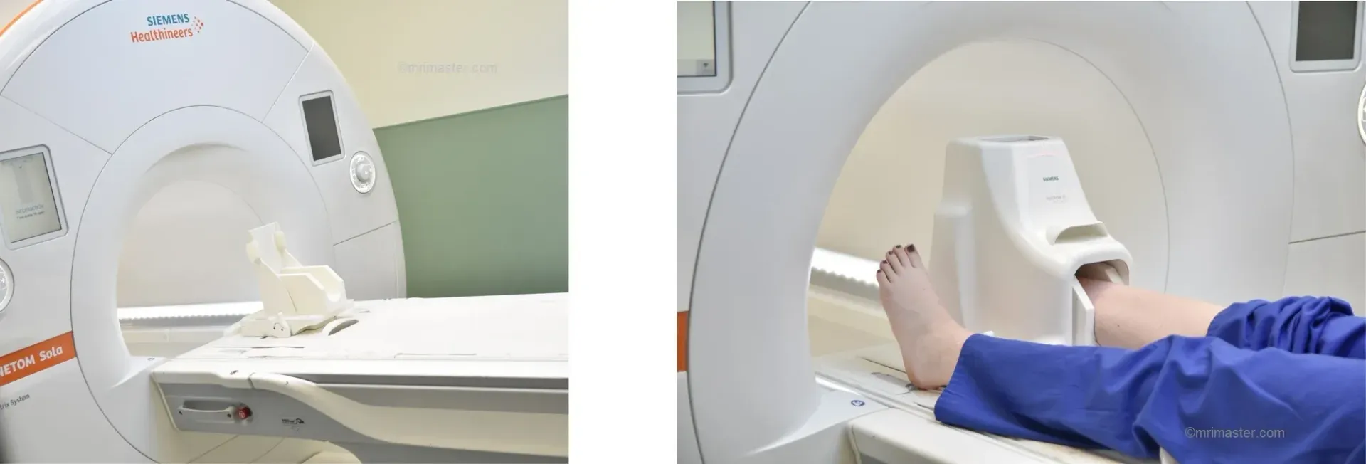
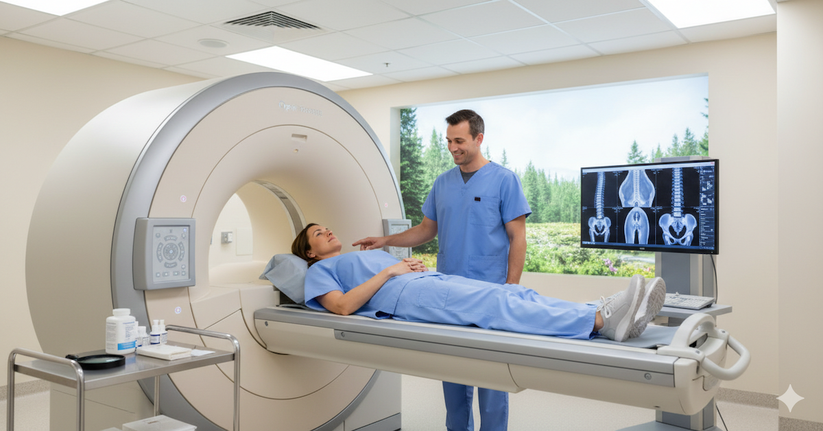
The procedure for obtaining an MRI foot scan is relatively straightforward. Before the scan, patients are positioned comfortably on the MRI machine, ensuring optimal imaging of the foot. It's essential to remove any metal objects or jewelry that could interfere with the scan. Once inside the MRI machine, patients may need to remain still for a period of time while the images are being captured.
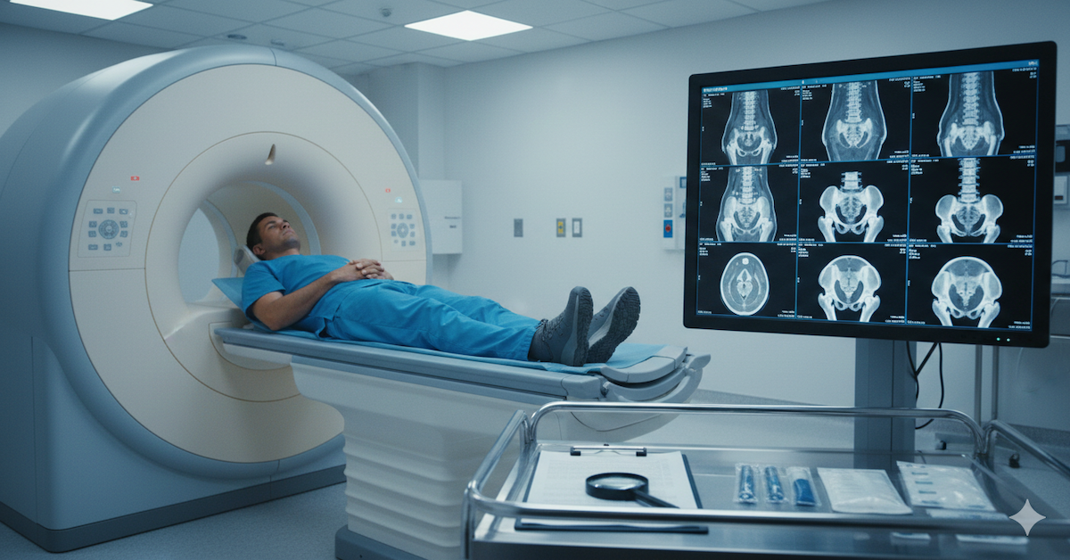
During the scan, patients may hear loud tapping or knocking noises, which is a normal part of the MRI process. While this can be unsettling for some, rest assured that the procedure is entirely safe and painless. After the scan is complete, the images are reviewed by a radiologist or orthopedic specialist, who will interpret the findings and discuss them with the patient.
Benefits of MRI Foot Scans
There are several benefits to undergoing an MRI foot scan. First and foremost, MRI scans are non-invasive, meaning there's no need for needles, incisions, or anesthesia. Additionally, MRI foot scans produce high-resolution images that offer unparalleled detail, allowing for a comprehensive evaluation of foot health. This level of precision is essential for accurate diagnosis and treatment planning.
Furthermore, MRI foot scans can be used to monitor disease progression or healing over time. Whether tracking the healing of a stress fracture or monitoring the progression of arthritis, MRI scans provide valuable information that can guide treatment decisions and interventions. Additionally, MRI foot scans can help identify underlying conditions that may be contributing to foot pain or dysfunction, allowing for targeted treatment approaches.
Applications and Considerations
MRI foot scans have a wide range of applications in clinical practice. From diagnosing sports injuries and degenerative conditions to evaluating congenital abnormalities and neuromuscular disorders, MRI scans play a vital role in foot health management. However, it's essential to consider factors such as cost and accessibility when recommending MRI foot scans to patients. While MRI technology is highly effective, it may not be readily available in all healthcare settings, and cost considerations may vary depending on insurance coverage and other factors.
Conclusion
In conclusion, MRI foot scans are a valuable diagnostic tool that provides essential insights into foot health and function. From identifying structural abnormalities to evaluating foot function and monitoring disease progression, MRI scans play a crucial role in the diagnosis and management of various foot conditions. If you're experiencing foot pain or discomfort, don't hesitate to discuss the possibility of an MRI foot scan with your healthcare provider. And remember, if you're in need of an MRI in the Deerfield area, consider Upright MRI of Deerfield for compassionate care and state-of-the-art imaging services.
SHARE THIS POST:
Leave a Comment:

The World's Most Patient-Friendly MRI. A comfortable, stress-free, and completely reliable MRI scan. We offer patients an open, upright, standup MRI experience that helps those who are claustrophobic and stress being in a confined area. Upright MRI of Deerfield is recognized as the world leader in open MRI innovation,
Our Recent Post


READ PATIENT TESTIMONIALS
Upright MRI of Deerfield.
Susan D.,
Highland Park, 39
I am going to tell everyone about your office! This was a great experience after I panicked in other MRI machines and had to leave. Thank you so much.

Judith B.,
Milwaukee, 61
I suffer from vertigo and other MRIs do not work. This was wonderful…absolutely NO discomfort at all. The MRI was so fast…I wanted to stay and watch the movie! Mumtaz was great. His humor really put me at ease. I’ve already recommended Upright MRI to friends.
Delores P.,
Glencoe, 55
Everything is so nice and professional with your place. I have been there a couple of times. My husband and I would not go anywhere else.

