
What Are the Benefits of MRI in Detecting Ehlers-Danlos Syndrome?
Ehlers-Danlos Syndrome, often shortened to EDS, is one of those conditions that can be hard to pin down. It doesn’t always follow a clear path. Symptoms vary widely, from flexible joints and chronic pain to more serious issues like fragile blood vessels or neurological complications. For people going through a long and confusing diagnostic journey, imaging tools like MRI can be incredibly helpful in putting the pieces together.
Understanding Why EDS Is Tough to Diagnose
EDS isn’t a single condition. It’s actually a group of connective tissue disorders, and each type brings its own unique challenges. Hypermobile EDS, which is the most common, often doesn’t show up on genetic tests. Classical and vascular types can involve more visible complications, like skin that bruises easily or blood vessels that are prone to tearing. The tricky part is that many symptoms overlap with other conditions. So even when a person knows something is off, getting a clear answer can take time. That’s where imaging, especially MRI, comes in as a valuable ally.
How MRI Helps in the Diagnostic Process
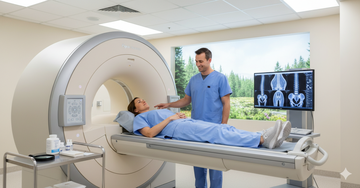
MRI, or magnetic resonance imaging, is one of the best ways to look at soft tissues inside the body without using radiation. It works by using magnetic fields to create detailed images of muscles, ligaments, joints, and internal organs. This makes it a great tool for exploring some of the issues that people with EDS often face but may not show during a regular physical exam. Since many symptoms involve soft tissue or neurological concerns, MRI provides a non-invasive way to look deeper and spot subtle signs that could be missed otherwise.
Digging Into the Benefits
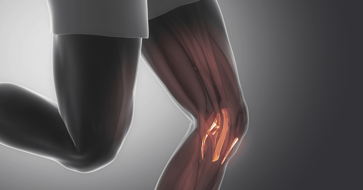
One of the biggest advantages of MRI in EDS cases is the ability to assess joints. People with EDS often experience frequent dislocations or joint subluxations, and these don’t always happen during an office visit. An MRI can catch joint damage, stretched ligaments, or cartilage wear that explains chronic pain or instability.
Spinal issues are another big area where MRI shines. Conditions like Chiari malformation, cervical instability, or tethered cord can all show up on an MRI scan. These are complications that might cause headaches, nerve pain, or coordination problems, and they’re not always visible without advanced imaging.
Soft tissue injuries can also fly under the radar. MRI can help find muscle tears, inflammation, or even herniated discs that contribute to daily discomfort. These kinds of issues are especially important to identify in people with EDS, as their tissues don’t heal the same way as others.
For those with vascular EDS, MRI can play a role in monitoring the health of blood vessels. In some cases, it can help detect aneurysms, vessel stretching, or other abnormalities before they lead to more serious problems. While it’s not a replacement for genetic confirmation or full vascular assessments, it adds a layer of safety and monitoring for high-risk individuals.
Neurological concerns are also common in some EDS types. MRI can help visualize cerebrospinal fluid leaks or craniocervical instability, which may contribute to dizziness, fatigue, or even fainting spells. When combined with clinical symptoms, these images can guide doctors toward a more complete understanding of what’s really going on.
Spotting Related Conditions with Confidence
Beyond just identifying EDS-related issues, MRI is useful for spotting conditions that often travel alongside it. Things like syringomyelia, where fluid-filled cysts develop in the spinal cord, or autonomic issues linked to nerve compression, can show up on scans. For patients who’ve spent years being misdiagnosed or told their symptoms are "all in their head," having visible proof on a scan can be a huge relief.
What MRI Can’t Do on Its Own
It’s important to understand that MRI doesn’t diagnose EDS directly. There’s no one image that says “yep, that’s EDS.” It’s more about using MRI to support the full picture. If someone has symptoms, a detailed history, and maybe even genetic markers or physical findings, the MRI can fill in the gaps. But on its own, it won’t tell the whole story.
Some MRI findings can also be subtle, and if the radiologist isn’t familiar with EDS-related complications, things might get missed. That’s why it’s helpful to work with providers who understand the condition or can consult with specialists who do.
A Team Approach Makes All the Difference
Diagnosing EDS properly often takes more than one expert. You might see a rheumatologist, neurologist, geneticist, or even a cardiologist depending on your symptoms. MRI works best when it’s part of that bigger team effort. It helps rule out other conditions, supports treatment planning, and gives visual confirmation of issues that are sometimes brushed off or misinterpreted.
Getting the right diagnosis isn’t just about putting a name to your symptoms. It’s about getting access to the care, therapies, and accommodations you need to improve your quality of life. And for many patients, MRI helps speed that process up.
Early Clues Lead to Better Outcomes
The sooner you can identify complications like spinal instability or vascular abnormalities, the better. Early intervention can help prevent further injury, manage pain more effectively, and reduce the chance of serious medical emergencies. MRI makes that kind of proactive care possible, especially when other tools fall short.
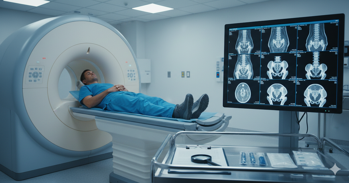
At Upright MRI of Deerfield, we offer imaging that’s designed to be comfortable and thorough for patients with chronic conditions like EDS. Our upright technology makes it easier to capture the way your body works in real-life positions, and our team understands that subtle findings can have a big impact. If you’re exploring a possible diagnosis or want a clearer view of what’s happening inside your body, we’re here to help with a caring, expert approach.
SHARE THIS POST:
Leave a Comment:

The World's Most Patient-Friendly MRI. A comfortable, stress-free, and completely reliable MRI scan. We offer patients an open, upright, standup MRI experience that helps those who are claustrophobic and stress being in a confined area. Upright MRI of Deerfield is recognized as the world leader in open MRI innovation,
Our Recent Post


READ PATIENT TESTIMONIALS
Upright MRI of Deerfield.
Susan D.,
Highland Park, 39
I am going to tell everyone about your office! This was a great experience after I panicked in other MRI machines and had to leave. Thank you so much.

Judith B.,
Milwaukee, 61
I suffer from vertigo and other MRIs do not work. This was wonderful…absolutely NO discomfort at all. The MRI was so fast…I wanted to stay and watch the movie! Mumtaz was great. His humor really put me at ease. I’ve already recommended Upright MRI to friends.
Delores P.,
Glencoe, 55
Everything is so nice and professional with your place. I have been there a couple of times. My husband and I would not go anywhere else.

