
What Foot Health Information Can MRI Scans Reveal?
Experiencing foot pain can be debilitating, affecting everything from daily activities to overall quality of life. For many, an MRI scan is a pivotal step toward uncovering the root causes of foot issues. Let's delve into how MRI scans provide essential insights into foot health, aiding in precise diagnosis and effective treatment.
Understanding the Structure of the Foot
Basic Anatomy of the Foot
The foot is a complex structure comprising bones, muscles, and ligaments that support body weight and enable movement. Common stress points in the foot often suffer from injuries due to overuse or improper foot mechanics, necessitating detailed imaging to diagnose problems accurately.
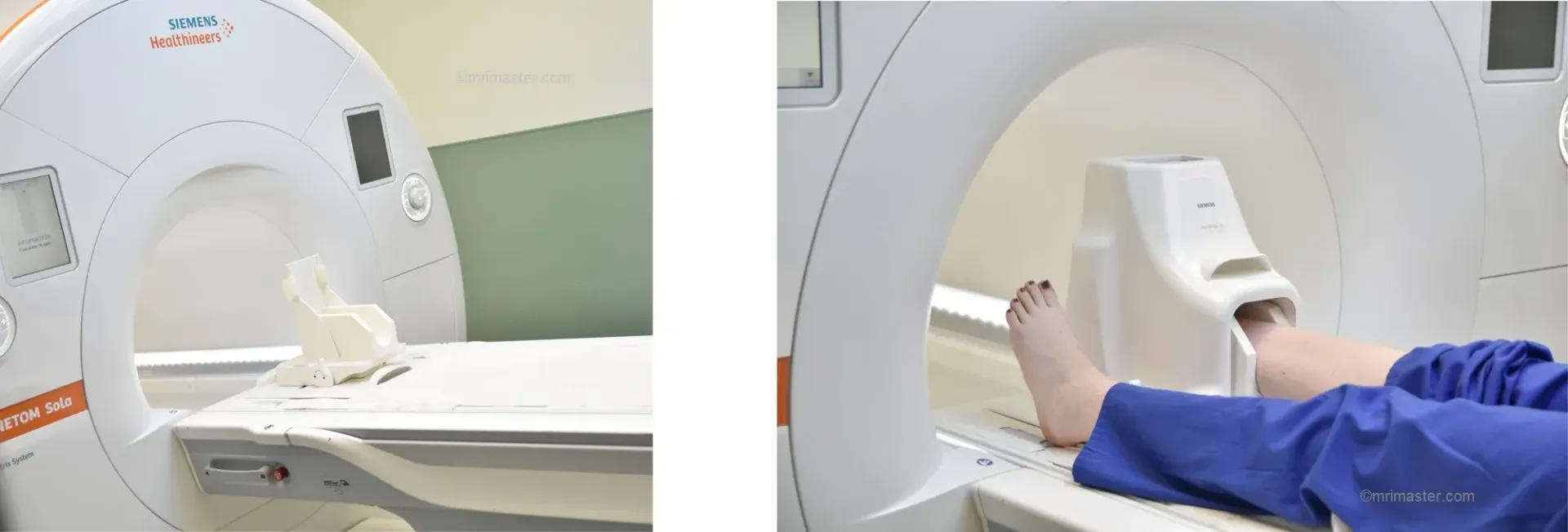
The Role of MRI in Foot Diagnostics
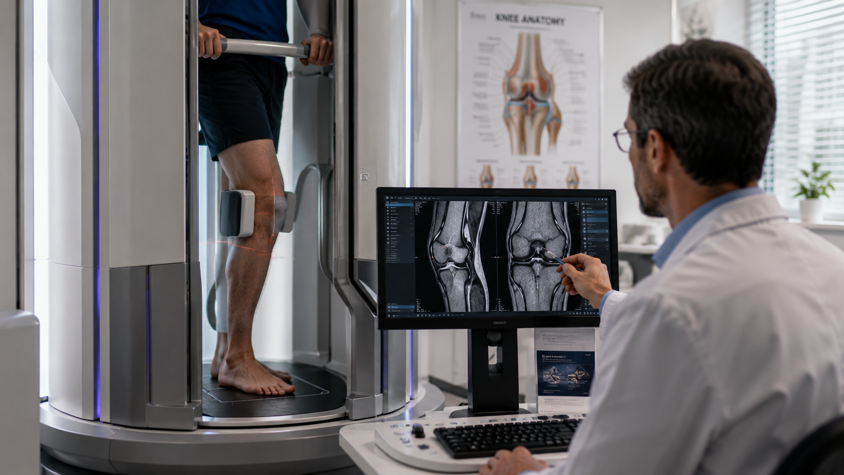
MRI scans offer a non-invasive method to obtain detailed images of the internal structures of the foot, surpassing other imaging techniques like X-rays, especially in visualizing soft tissue. This capability is crucial for diagnosing a wide range of foot conditions.
Key Insights Provided by MRI Scans
Soft Tissue Injuries
Tendonitis and Tendon Tears: MRI scans excel in identifying changes in tendons, such as tears or inflammation, which are common in athletes or individuals with repetitive strain injuries. For instance, MRI can clearly show the severity of an Achilles tendon tear, guiding the treatment approach, whether surgical or non-surgical.
Ligament Strains and Tears: Ligaments in the foot, when damaged, can lead to instability and pain. MRI can pinpoint the specific ligaments affected and the extent of the injury, crucial for targeted treatments and rehabilitation strategies.
Bone Health
Stress Fractures: Unlike X-rays, which might miss early signs of stress fractures, MRI scans can detect these subtle fractures and even bone marrow edema that often accompanies or signals the onset of more significant bone injuries. This early detection is vital for preventing further damage.
Bone Marrow Edema: MRI is sensitive enough to detect bone marrow edema—an indicator of trauma or degenerative changes within the bone—before more significant problems arise, allowing for early intervention.
Joint Conditions
Arthritis Detection: For those suffering from joint pain in the feet, MRI scans can detect the early stages of arthritis, showing areas of cartilage wear and joint inflammation. This early diagnosis is essential for managing symptoms and slowing the disease's progression.
SynovitisIn cases of joint pain and swelling, MRI helps differentiate synovitis (inflammation of the joint lining) from other conditions, ensuring that treatment is appropriately targeted to reduce inflammation and restore function.
Nerve Conditions
Morton’s Neuroma: This painful condition involves the thickening of nerve tissue between toes, commonly detected by MRI. The scan provides clear images of the affected nerves, helping in formulating a precise treatment plan.
Tarsal Tunnel Syndrome: MRI scans are instrumental in diagnosing Tarsal Tunnel Syndrome, a condition caused by the compression of nerves within the tarsal tunnel of the ankle. The detailed images help confirm the diagnosis and guide subsequent interventions.
Challenges and Limitations of MRI for Foot Health
Accessibility and Cost
While MRI offers unparalleled imaging capabilities, access to high-quality MRI can be limited, and the cost can be prohibitive for some patients. It's important for patients to weigh these factors and explore all available imaging options.
Patient Considerations
Not everyone is a candidate for an MRI. For instance, patients with certain types of implants or those who experience severe claustrophobia might face challenges with standard MRI machines. However, advancements such as open and upright MRI machines, like those offered at Upright MRI of Deerfield, are making scans more accessible and comfortable for all patients.
Conclusion
MRI scans are a powerful tool in diagnosing various foot conditions, providing detailed images that are crucial for accurate diagnosis and effective treatment. By understanding the capabilities and limitations of MRI technology, patients can better navigate their healthcare options.
If you're experiencing foot pain or other related issues and think an MRI might be the right choice for you, consider reaching out to Upright MRI of Deerfield. Our advanced imaging solutions and patient-centric approach ensure you receive the best care and diagnostics available. Contact us today to learn more about how we can help you step back into comfort and health.
SHARE THIS POST:
Leave a Comment:

The World's Most Patient-Friendly MRI. A comfortable, stress-free, and completely reliable MRI scan. We offer patients an open, upright, standup MRI experience that helps those who are claustrophobic and stress being in a confined area. Upright MRI of Deerfield is recognized as the world leader in open MRI innovation,
Our Recent Post


READ PATIENT TESTIMONIALS
Upright MRI of Deerfield.
Susan D.,
Highland Park, 39
I am going to tell everyone about your office! This was a great experience after I panicked in other MRI machines and had to leave. Thank you so much.

Judith B.,
Milwaukee, 61
I suffer from vertigo and other MRIs do not work. This was wonderful…absolutely NO discomfort at all. The MRI was so fast…I wanted to stay and watch the movie! Mumtaz was great. His humor really put me at ease. I’ve already recommended Upright MRI to friends.
Delores P.,
Glencoe, 55
Everything is so nice and professional with your place. I have been there a couple of times. My husband and I would not go anywhere else.

