
How Do MRI Scans Contribute to Precision Diagnosis of Shoulder Conditions?
Experiencing shoulder pain can be frustrating, especially when the cause remains unclear. For many dealing with such discomfort, an MRI scan often becomes a crucial step towards finding relief. This advanced imaging technology plays a pivotal role in diagnosing various shoulder conditions accurately, guiding effective treatment plans. Here’s a closer look at how MRI scans help pinpoint shoulder issues with remarkable precision.
Understanding Shoulder Anatomy and Common Conditions
Basic Anatomy of the Shoulder
The shoulder is a complex assembly of structures including bones, tendons, ligaments, and muscles. These components collaborate to provide extensive mobility, making the shoulder prone to various injuries and conditions.
Common Shoulder Conditions
Shoulder issues range from rotator cuff tears and impingement syndromes to arthritis. Each condition presents with overlapping symptoms such as pain and restricted movement, posing challenges in diagnosis. Precise imaging is essential to differentiate these conditions and determine the best course of treatment.
Role of MRI in Diagnosing Shoulder Conditions
Advantages of MRI Scans
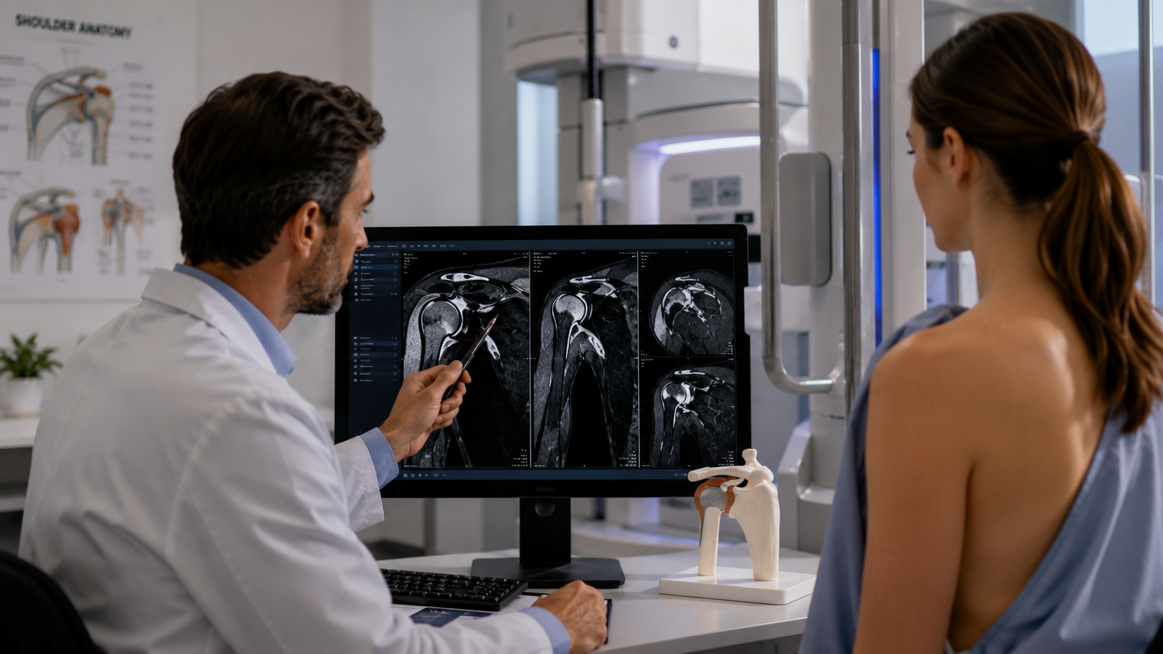
MRI scans stand out for their ability to produce high-resolution images of the shoulder’s soft tissues, bones, and cartilage without invasive procedures. This detail is invaluable in medical diagnostics, offering a clear view of the area without any surgical risks.
MRI Technology Explained
MRI uses magnetic fields and radio waves to create detailed pictures of the internal structures of the shoulder. This technique is particularly beneficial for examining soft tissues, making it superior for detecting subtle or hidden abnormalities.
Types of MRI for Shoulder Diagnosis
Standard MRI provides comprehensive images, but certain conditions may require specialized techniques:
- MR Arthrography involves injecting a contrast dye into the joint to provide a clearer view of its internal structure, useful for complex diagnoses.
- Functional MRI assesses the shoulder in various positions or states of use, helping to understand the mechanics behind the symptoms.
MRI Diagnosis of Specific Shoulder Conditions
Rotator Cuff Tears
MRI scans excel in identifying and assessing the severity of rotator cuff tears. They show both partial and complete tears, offering crucial information that impacts treatment decisions, from conservative management with physical therapy to surgical repair.
Impingement Syndrome and Bursitis
For conditions like impingement syndrome and bursitis, MRI effectively visualizes inflammation and swelling. Detecting these early can prevent further complications and tailor treatments such as anti-inflammatory medications or injections to specific needs.
Arthritis and Degenerative Conditions
In cases of arthritis, MRI scans detect early signs of joint wear and cartilage loss. They provide a baseline for monitoring disease progression and adjusting treatments over time, a crucial aspect in managing chronic conditions.
Challenges and Limitations of MRI Scans
Limitations in Specific Populations
Not everyone can undergo a standard MRI. Patients with certain metal implants, pacemakers, or other conditions may face restrictions due to safety concerns with magnetic fields.
Advances in MRI Technology
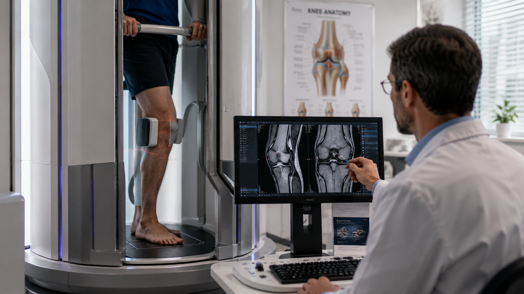
To accommodate more patients, including those who are claustrophobic or cannot lie flat due to pain, advancements such as open and upright MRI machines have been developed. These alternatives offer more comfort without compromising the quality of diagnostics, broadening the accessibility of precise imaging.
Conclusion
MRI scans are indispensable in the precise diagnosis of shoulder conditions. They provide detailed images that are crucial for accurately identifying and treating various disorders. As technology advances, the capabilities of MRI in diagnosing and managing shoulder problems continue to expand, offering hope and relief to those suffering from chronic pain and mobility issues.
At Upright MRI of Deerfield, we understand the critical role of accurate diagnostics in treating shoulder conditions. Our state-of-the-art MRI technology, including our upright MRI capabilities, ensures that all patients receive the highest standard of care and comfort during their imaging procedures. If you're struggling with shoulder pain and need precise diagnostics, contact us today to see how we can help you get back to your best health.
SHARE THIS POST:
Leave a Comment:

The World's Most Patient-Friendly MRI. A comfortable, stress-free, and completely reliable MRI scan. We offer patients an open, upright, standup MRI experience that helps those who are claustrophobic and stress being in a confined area. Upright MRI of Deerfield is recognized as the world leader in open MRI innovation,
Our Recent Post


READ PATIENT TESTIMONIALS
Upright MRI of Deerfield.
Susan D.,
Highland Park, 39
I am going to tell everyone about your office! This was a great experience after I panicked in other MRI machines and had to leave. Thank you so much.

Judith B.,
Milwaukee, 61
I suffer from vertigo and other MRIs do not work. This was wonderful…absolutely NO discomfort at all. The MRI was so fast…I wanted to stay and watch the movie! Mumtaz was great. His humor really put me at ease. I’ve already recommended Upright MRI to friends.
Delores P.,
Glencoe, 55
Everything is so nice and professional with your place. I have been there a couple of times. My husband and I would not go anywhere else.

