
The Foot in Focus: Understanding MRI Foot Scans
Our feet, elaborate in layout and typically underappreciated, are the unhonored architects of our physical security. When foot disorders strike, they can resound through our whole bone and joint system, impacting our gait, posture, and overall well-being. This is where the precision of MRI foot checks enters into play, providing a lens into the complicated makeup of the feet, allowing precise medical diagnoses and customized therapy plans.
The Essentials of MRI Foot Scans
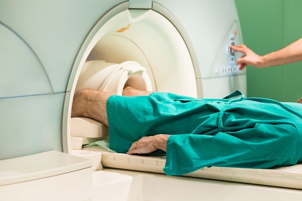
MRI, the master of contemporary clinical imaging, utilizes a harmony of magnetic fields and radio waves to unravel the secrets of foot composition. It resembles peeling back the layers of a complex tapestry, exposing cross-sectional pictures that far go beyond the capacities of traditional X-rays and CT scans. The remarkable soft tissue comparison offered by MRI is specifically critical in foot diagnostics, where a comprehensive examination of bones, joints, tendons, and ligaments is vital.
Applications of MRI in Assessing Foot Makeup
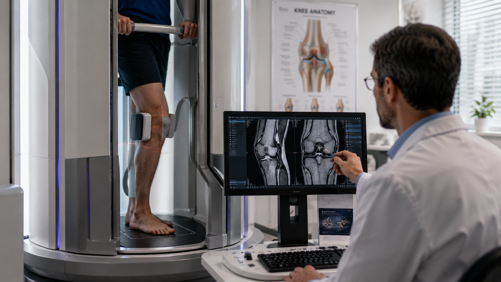
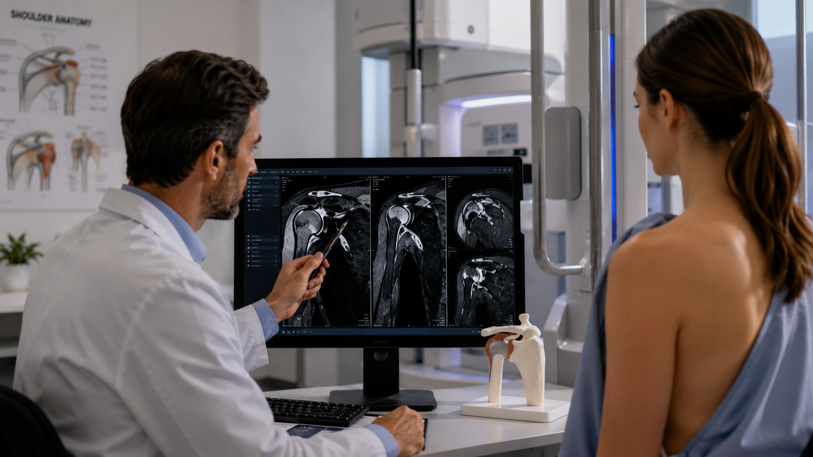
Photo this: a high-resolution picture recording the delicate dance between bones and soft tissues in the foot. MRI excels in bone and joint imaging, enabling clinicians to find cracks, joint inflammation, and various other orthopedic conditions with unequalled precision. Furthermore, soft tissue assessment is a strong suit of MRI, making it possible for the recognition of injuries, splits, and inflammation in tendons and tendons. It belongs to having a front-row seat to the elaborate ballet of foot composition.
MRI Foot Checks for Certain Foot Conditions
Zooming in on details foot problems, MRI becomes a diagnostic giant. Take Plantar Fasciitis, for example- MRI introduces the swelling of the plantar fascia, leading treatment alternatives and rehabilitation approaches. Stress fractures, usually elusive in standard X-rays, come to light with the comprehensive imaging capacities of MRI, helping with very early treatment and preventing potential complications. Tendon problems, such as Achilles tendonitis, are scrutinized with accuracy, assisting in the tailoring of effective therapy plans.
Benefits of MRI Foot Checks
Past its analysis capabilities, the attraction of MRI foot checks depend on their patient-friendly nature. Non-invasive and devoid of ionizing radiation, MRI guarantees the safety of people. The high-resolution imaging not just supplies a detailed medical diagnosis however also provides a thorough view of both bones and soft cells in a solitary check. It's an all natural approach to foot health that establishes MRI apart as a diagnostic ally.
Considerations and Individual Experience
Getting in the MRI world may stimulate problems, specifically when it pertains to claustrophobia. However, innovations in open MRI systems resolve this, giving a much more comfortable experience for patients undergoing foot scans. Individual participation is paramount for ideal check quality, and the concentrate on enhancing the general experience underscores the dedication of doctor to making foot imaging as seamless as possible.
Future Fads in MRI Foot Imaging
The future of MRI foot imaging is as exciting as it is promising. The combination of artificial intelligence is positioned to raise picture evaluation, making diagnostics a lot more accurate and effective. As innovation evolves, the focus is not just on progressing diagnostic capabilities but also on boosting the general client experience, guaranteeing access and convenience in the world of foot wellness diagnostics.
Verdict
In concluding our exploration into the world of MRI foot scans, it appears that this innovation is not nearly diagnostics; it's about accuracy, empathy, and the elaborate dance of innovation with human composition. The feet, commonly overlooked, discover a spotlight in the detailed images offered by MRI, paving the way for a much deeper understanding of foot wellness and more reliable therapy approaches.
As we look into the ins and outs of foot health, Upright MRI of Deerfield stands at the center of this clinical change. Our commitment to leveraging sophisticated technology underscores our commitment to progressing foot health and wellness and ensuring the wellness of our individuals. Discover the complexities of foot health and wellness with us-- where precision meets compassion, and innovation fulfils care.
SHARE THIS POST:
Leave a Comment:

The World's Most Patient-Friendly MRI. A comfortable, stress-free, and completely reliable MRI scan. We offer patients an open, upright, standup MRI experience that helps those who are claustrophobic and stress being in a confined area. Upright MRI of Deerfield is recognized as the world leader in open MRI innovation,
Our Recent Post


READ PATIENT TESTIMONIALS
Upright MRI of Deerfield.
Susan D.,
Highland Park, 39
I am going to tell everyone about your office! This was a great experience after I panicked in other MRI machines and had to leave. Thank you so much.

Judith B.,
Milwaukee, 61
I suffer from vertigo and other MRIs do not work. This was wonderful…absolutely NO discomfort at all. The MRI was so fast…I wanted to stay and watch the movie! Mumtaz was great. His humor really put me at ease. I’ve already recommended Upright MRI to friends.
Delores P.,
Glencoe, 55
Everything is so nice and professional with your place. I have been there a couple of times. My husband and I would not go anywhere else.

