
How MRIs Save Lives With Early Cancer Detection
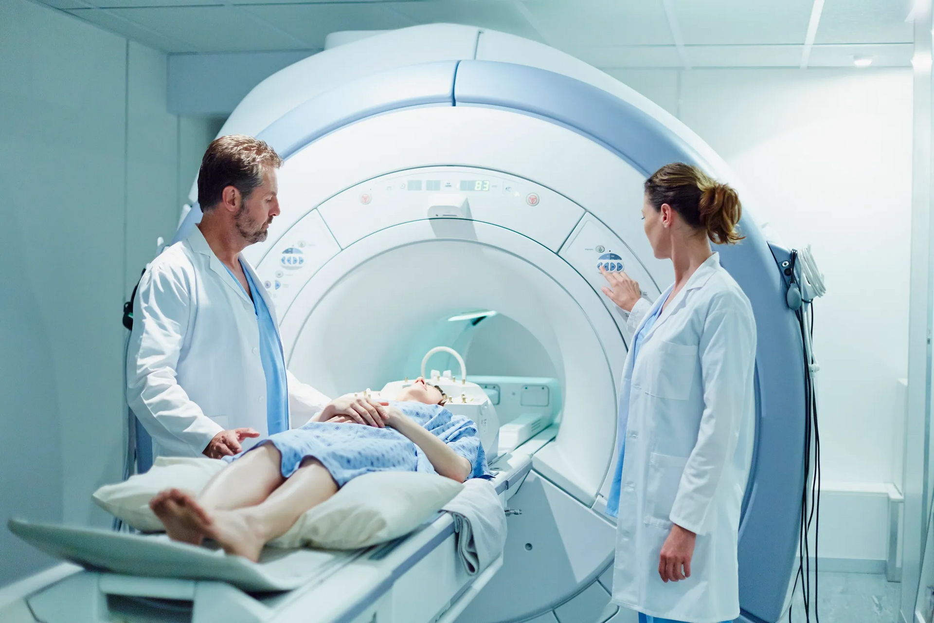
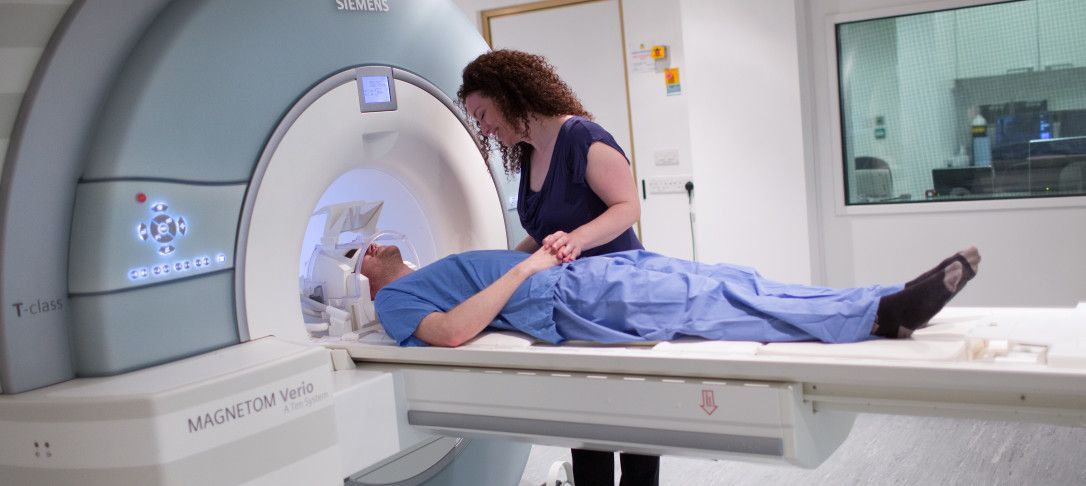
MRI, or magnetic resonance imaging, exams are lifesaving procedures in the fight against brain and spinal cord cancer. Doctors use MRI exams to detect cancer early, before it has metastasized. Cancer is much more treatable if doctors find the tumors in the beginning stages. Because of MRIs, families every year watch their loved ones recover from potentially life-ending illnesses.
What Do MRIs Show?
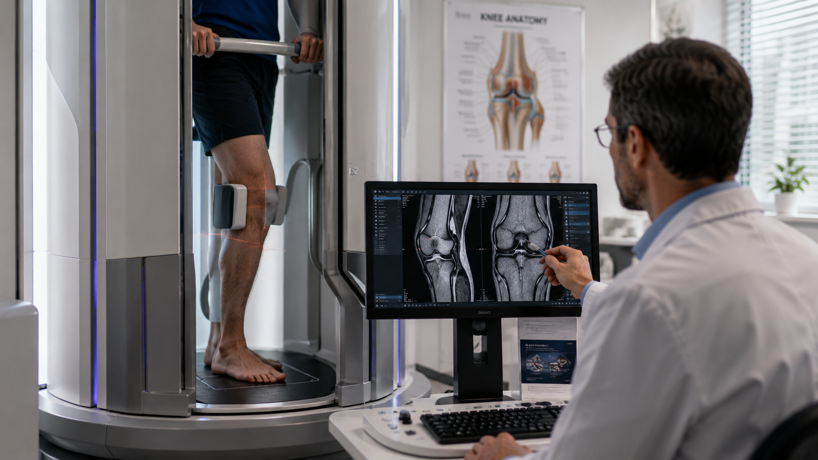
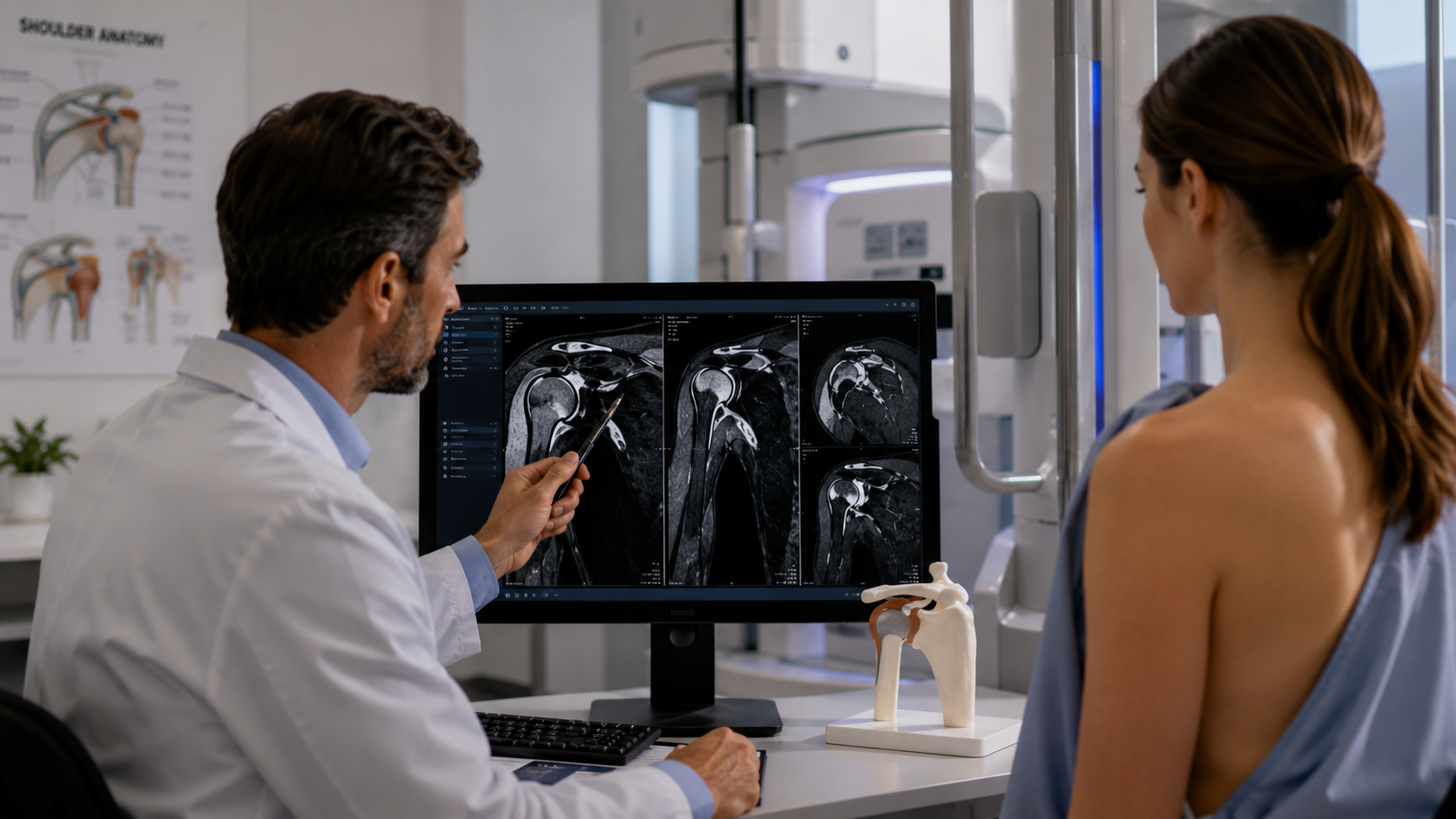
Using MRIs, doctors can develop cross-section images of a person’s insides. In other words, MRIs create images in “slices.” They explore bodies through multiple angles. When doctors look at MRI pictures, they see a person from the side, front, and above. MRIs can detect parts of a person that are sometimes difficult to uncover using other tests. This is one of the main reasons why MRIs are a favored imaging technique for some tumors.
What Cancers Can MRIs See?
MRIs are extremely adept at seeing certain kinds of tumors. An MRI with contrasting dye is the preferred method for detecting spinal cord and brain tumors. MRIs can detect these tumors early in their development, before they have metastasized. Early detection is very important for cancer treatment.
When cancer metastasizes, it spreads to other parts of the body. It’s a sign that the cancer has become more serious, more deadly. In many cases, there is little a doctor can do after the cancer has grown into multiple parts of the body. One of the great things about MRIs is they can detect tumors early. Early detection allows doctors to treat cancer in a timely manner.
Why Early Detection Is Important
There is no doubt that cancer is most treatable when we detect it early. Too often doctors find tumors when they have already developed into other areas and, as a result, are unable to make the quick decisions that save lives. Using MRIs, we detect tumors of the brain and spinal cord early enough to make a difference. Doctors can use the information gathered from MRIs to determine specialized therapies. This is why it’s very important to get tested for cancer. Unfortunately, many people don’t detect their cancers soon enough.
Why People Put Off Cancer Tests
If you talk to doctors about the most upsetting parts of the job, they will likely reference the people they could not save because their illnesses were not detected soon enough. Death is always a tragedy, but death that could have been avoided is especially painful. There are many reasons people put off MRIs that detect cancers, including:
- It can be expensive. Especially for underinsured people, medical expenses add up. Many people avoid tests because they don’t want to pay the bill. But, in truth, there are few things more worth your money than your health. Don’t let your cancer go undetected just because you didn’t want to pay.
- Bad news can be frightening. Some people avoid medical examinations like MRIs because they are afraid the tests could uncover bad news. People are so nervous about what the MRI could reveal that they decide to skip the procedure altogether. Yes, MRIs can be scary. No one wants to learn that they have cancer. But if a
doctor recommends you undergo an MRI, it doesn’t automatically mean bad news. They just want to take the precaution. Also, if you do have cancer, ignoring the problem will not offer a solution. It’s better to get tested and face the truth.
- People are incorrectly nervous about the radiation. MRI machines work their magic by creating a magnetic field around the person being studied. During the process, people are exposed to radio waves. Some patients have the misconception that this technology is dangerous. But that is incorrect. An MRI’s radiation is relatively harmless. Do not let a baseless fear about radiation get in the way of learning vital medical information.
What Doctors Do When They Spot a Brain Tumor
Doctors can spot tumors using MRIs, but they don’t always immediately know what kind of tumor it is. Is it benign or malignant? A malignant tumor is a cancerous growth, one that has the potential to metastasize and kill a person. A benign tumor is a growth that will not metastasize. Doctors can determine for certain if the tumor is benign or malignant by performing a biopsy. A biopsy is a procedure wherein a doctor removes part or all of the tumor, sometimes to determine if it is cancerous and sometimes to get rid of a dangerous growth. For brain tumors, there are two kinds of biopsies:
Stereotactic (needle) biopsy. Doctors normally use a stereotactic, or needle, biopsy when it is too risky to remove the tumor entirely. People who have tumors deep in their brain, so deep that cutting through the brain to remove it would be problematic, will often receive this kind of biopsy. For stereotactic biopsies, doctors will make an incision into the scalp and drill a tiny hole into the skull. Leveraging image-guidance systems, they remove a piece of tissue for study. Afterward, a pathologist reviews the sample to determine if it’s cancerous. Based on that diagnosis, the doctor will decide what kind of treatment they should use to combat the disease.
Surgical or open biopsy (craniotomy). For surgical or open biopsies, doctors can remove most or all of the tumor outright. These are for tumors that are not in hard-to-reach positions. Doctors utilize a procedure called a craniotomy. They start by removing a small part of the tumor and analyzing it while the patient is still on the operating table. This will help guide treatment and determine if they should remove the rest of the tumor. If it’s too risky to remove the entire tumor, doctors may just take part of it in a process called debulking.
Conclusion
Cancer is a horrifying thing. No one wants to be told that a dangerous growth has invaded and attacked their body. But it’s absolutely important that people who may have cancer seek out procedures that let them know one way or the other. For brain and spinal cord tumors, there is no better detection device than the MRI. These machines can detect a tumor early enough to make a difference. When it comes to cancer, the most important thing is early detection.
SHARE THIS POST:
Leave a Comment:

The World's Most Patient-Friendly MRI. A comfortable, stress-free, and completely reliable MRI scan. We offer patients an open, upright, standup MRI experience that helps those who are claustrophobic and stress being in a confined area. Upright MRI of Deerfield is recognized as the world leader in open MRI innovation,
Our Recent Post


READ PATIENT TESTIMONIALS
Upright MRI of Deerfield.
Susan D.,
Highland Park, 39
I am going to tell everyone about your office! This was a great experience after I panicked in other MRI machines and had to leave. Thank you so much.

Judith B.,
Milwaukee, 61
I suffer from vertigo and other MRIs do not work. This was wonderful…absolutely NO discomfort at all. The MRI was so fast…I wanted to stay and watch the movie! Mumtaz was great. His humor really put me at ease. I’ve already recommended Upright MRI to friends.
Delores P.,
Glencoe, 55
Everything is so nice and professional with your place. I have been there a couple of times. My husband and I would not go anywhere else.

