
How Does Neutral vs Flexion MRI Change Diagnosis?
The human spine is not a static structure. It bends, extends, rotates, and bears load throughout every hour of waking life. The symptoms that bring most patients to imaging, pain, stiffness, radiating nerve symptoms, and loss of function, are almost always experienced during movement or under load rather than while lying flat and perfectly still. Yet the overwhelming majority of spinal MRI studies are performed with the patient in exactly that position: lying supine in a neutral posture, removed from all the mechanical conditions that produce symptoms.
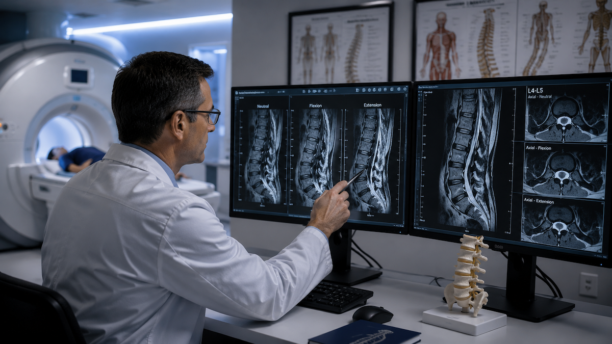
This mismatch between how the spine is imaged and how symptoms actually present is the central problem that neutral versus flexion MRI comparisons reveal so clearly. When the same spine is imaged in neutral and in flexion, the findings are often strikingly different. Canal dimensions change. Disc geometry shifts. Ligamentous laxity becomes measurable. Foraminal areas narrow or widen. The spine that appeared unremarkable in neutral may display the structural basis of significant symptoms in flexion.
Quick Answer: A neutral MRI images the spine at rest with no mechanical load and no positional stress. A flexion MRI images the spine in forward bending, under load or in the upright seated position. The two studies routinely produce different findings because the anatomy changes with position. Neutral imaging underestimates or misses dynamic stenosis, positional disc herniation, segmental instability, and ligamentous laxity that only become apparent when the spine is placed under the functional stress that produces symptoms. Flexion imaging captures the spine in the condition most relevant to the patient's actual experience.
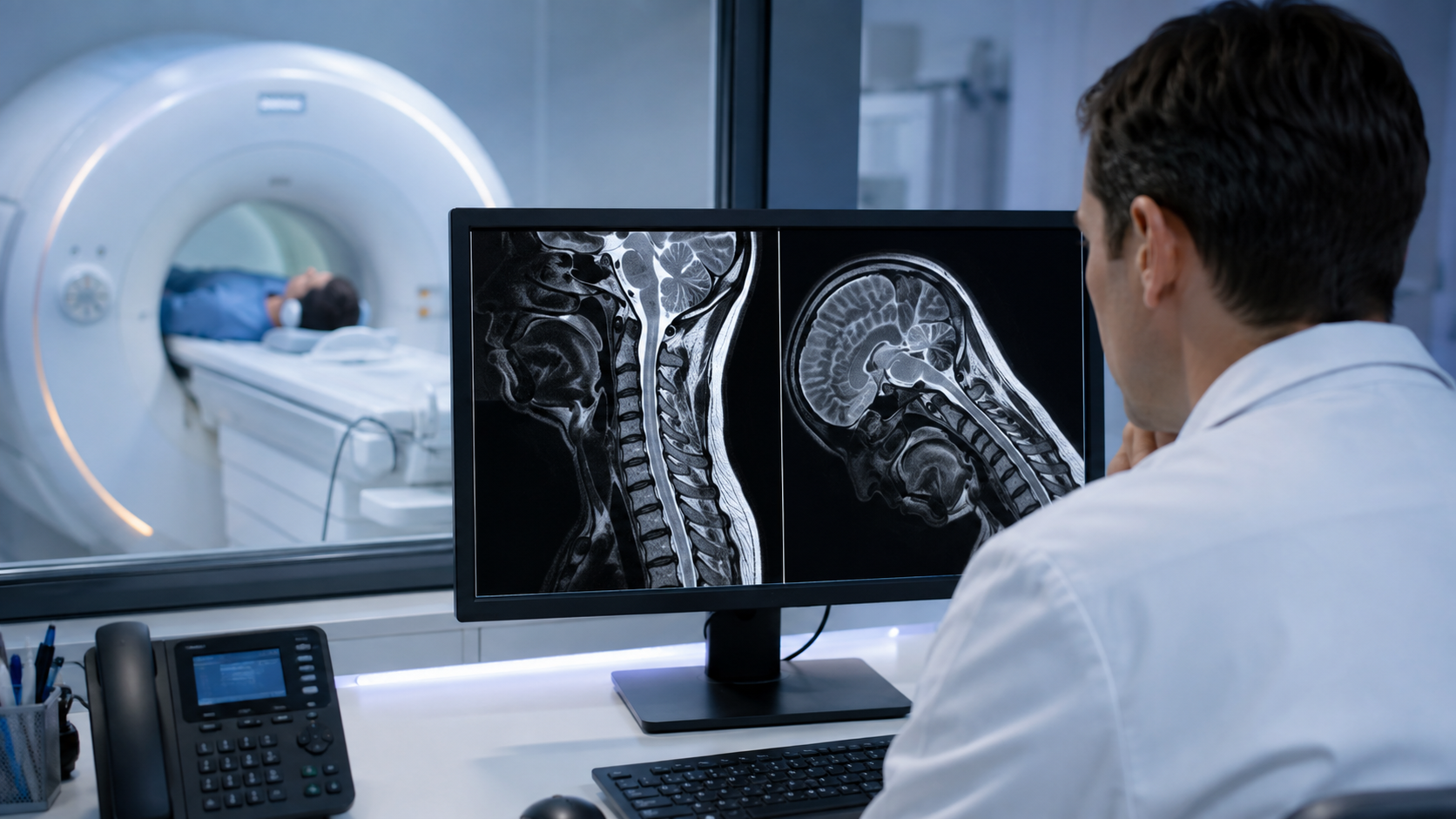
What Changes in the Spine During Flexion
Several structural changes occur simultaneously when the spine moves from neutral to flexion. In the lumbar spine, forward bending opens the posterior elements, widens the intervertebral foramina, and reduces the anterior disc height while increasing the posterior disc height.
In the cervical spine, flexion can increase the AP diameter of the spinal canal in some configurations while creating new compressive forces at specific segments. At any spinal level, ligamentum flavum changes its mechanical state during movement in ways that directly affect the space available to neural structures.
At segments with underlying instability, these changes are amplified beyond what occurs in a healthy spine. An unstable segment that shows 2mm of anterior translation in neutral may show 7mm in flexion. A disc that appears minimally herniated in neutral may show significant posterior protrusion when the spine is loaded in flexion. A foraminal area that measures adequate in supine imaging may be critically narrowed in the position the patient actually adopts when their symptoms are worst.
The clinical significance of these positional changes is why flexion and extension MRI is considered essential rather than supplementary for specific diagnostic questions, particularly those involving instability, dynamic stenosis, and symptoms that are clearly position-dependent.
Dynamic Stenosis: The Classic Example
Spinal stenosis is the narrowing of the spinal canal or neural foramina to the point where neural structures are compressed. In a neutral supine MRI, the canal and foramina are at their most decompressed state: the posterior ligamentous structures are relatively taut and out of the canal, the disc is unloaded, and the facet joints are at rest. This is when stenosis appears at its least severe.
In flexion and under gravitational load, the same space may be significantly smaller. The posterior disc bulges further into the canal as axial load is applied. The ligamentum flavum buckles inward as the posterior elements come together. Facet effusions can expand under loading. The cumulative effect is that a canal that measured adequate in supine imaging becomes significantly narrowed when the patient stands, walks, or performs the activities that produce their symptoms.
This dynamic behaviour explains why supine MRI limitations are most pronounced for patients whose symptoms occur specifically with activity: the imaging is performed in the one position where the problematic narrowing is at its least apparent.
Segmental Instability: Only Visible Under Stress
Segmental instability, the abnormal movement of one vertebral segment relative to the adjacent one beyond the normal range of motion, is by definition a dynamic finding. It exists in the movement of the spine, not in its resting state.
A neutral MRI can show the structural conditions that predispose to instability, such as disc degeneration, facet arthropathy, or ligamentous thickening, but it cannot measure the actual movement that constitutes instability because no movement is occurring during the scan.
Flexion-extension imaging quantifies this movement directly. By comparing the position of vertebral bodies relative to each other in maximum flexion and maximum extension, the amount of translational or angular movement at each level can be measured. Segments where this movement exceeds normal parameters are identified as unstable, which carries direct implications for treatment decisions including whether stabilisation is needed and how many levels need to be addressed.
In the lumbar region, lumbar flexion MRI findings of dynamic instability are among the most clinically significant positional discoveries, particularly in patients who have been told their neutral MRI is normal or near-normal despite significant functional limitation.
Disc Pathology That Changes With Position
Not all disc herniations are fixed structural events. Some disc pathology is genuinely positional: the disc material moves or the herniation changes in size and configuration with spinal positioning. A disc that shows a minimal central protrusion in neutral may show a clinically significant lateral herniation into the neural foramen when the spine is loaded in flexion. The reverse can also occur: some herniations are more prominent in extension than in flexion.
For patients whose radicular symptoms, the shooting pain, numbness, or weakness that travel into the limbs, are provoked only in specific positions, this positional disc behaviour is the most likely structural explanation. Imaging in the symptom-provoking position captures the herniation in its most clinically relevant configuration, which is the information actually needed to plan treatment.
Ligamentous Laxity and Soft Tissue Changes
The posterior longitudinal ligament, the ligamentum flavum, and the facet joint capsules all change their mechanical state during spinal movement. In patients with ligamentous laxity, whether from connective tissue disorders, prior trauma, or chronic degenerative loosening, these structures allow more movement than they should and may permit neural compression under loading that would not occur in a healthy spine.
This is particularly significant in the cervical spine, where the consequences of cord compression can be serious. Cervical ligamentous instability that is absent on neutral imaging may produce cord flattening or myelopathic signal change in flexion that represents a significant finding with direct implications for management.
The range of conditions needing positional MRI includes connective tissue disorders, post-traumatic instability, failed back surgery syndrome with recurrent symptoms, and any presentation where the clinical symptoms are significantly more severe than the neutral imaging findings would predict.
How Flexion Findings Change Clinical Management
The diagnostic difference between neutral and flexion imaging translates directly into different clinical decisions. A patient whose neutral MRI shows mild degenerative changes may receive conservative management indefinitely without a clear structural target. The same patient, imaged in flexion, may show dynamic stenosis at a specific level, segmental instability at another, and a positional disc herniation at a third. Each of these findings has a specific treatment pathway that the neutral imaging did not open.
For surgical planning in particular, identifying all levels of significant dynamic pathology before an operation prevents the complication of addressing some levels while leaving others that will require further surgery. For non-surgical management, a specific positional diagnosis allows targeted injection, specific physiotherapy protocols, and activity modification advice that is grounded in what the imaging actually shows rather than what the neutral study suggested.
Referring clinicians who want to discuss specific imaging protocols for patients with position-dependent symptoms will find detailed guidance on the physician referral page at Upright MRI of Deerfield, including how to specify the positions most relevant to each clinical scenario.
The principle that weight-bearing MRI provides a more functionally relevant picture of spinal pathology applies equally to the neutral versus flexion comparison: the closer the imaging conditions are to the conditions that produce symptoms, the more clinically useful the findings become.
Frequently Asked Questions
Can both neutral and flexion MRI be performed in the same appointment?
Yes. An upright positional MRI system allows the spine to be imaged in multiple positions within a single session, including neutral, flexion, and extension. The comparative data from a single session is more reliable than comparing studies performed at different times because patient positioning and the time of day can affect spinal measurements.
Does a normal neutral MRI mean nothing is wrong with the spine?
No. A normal neutral MRI means the spine appears structurally within normal limits in the supine unloaded position. It does not rule out positional or dynamic pathology that is only present under load or in specific positions. For patients with symptoms that are clearly activity-dependent and a normal neutral MRI, positional imaging is a logical next step.
What conditions benefit most from comparing neutral and flexion MRI?
Spondylolisthesis where the degree of slip changes with position, cervical and lumbar instability, dynamic foraminal stenosis, positional disc herniation, and symptoms that are significantly worse in specific positions all benefit from the neutral-to-flexion comparison. The comparison provides the dynamic dimension that neutral imaging alone cannot supply.
Will my insurance cover a flexion MRI after a normal neutral scan?
Coverage depends on the insurer and the clinical justification. Demonstrating that the neutral scan was performed, that symptoms persist and are position-dependent, and providing a clinical rationale for why positional imaging is likely to add diagnostic value supports the case for coverage. Some insurers require a physician's letter of medical necessity.
How do I request a flexion MRI after receiving a normal neutral scan report?
Discuss the option with your referring physician and ask them to specify flexion and extension sequences in the referral. Contacting a centre that performs upright positional imaging directly is also an option, as they can advise on what positions are most relevant based on your symptom description and can coordinate with your physician.
The Bottom Line
A neutral MRI and a flexion MRI of the same spine frequently produce meaningfully different findings because the spine's anatomy changes with position. Dynamic stenosis, segmental instability, positional disc herniation, and ligamentous laxity are all categories of pathology that neutral imaging underestimates or misses entirely. For patients whose symptoms are position-dependent, the flexion study is often the one that provides the clinically useful answer.
Upright MRI of Deerfield performs neutral, flexion, extension, and weight-bearing spinal imaging in a single appointment. If a neutral scan has not explained your symptoms, contact the team to discuss whether positional imaging would provide the missing diagnostic information.
SHARE THIS POST:
Leave a Comment:

The World's Most Patient-Friendly MRI. A comfortable, stress-free, and completely reliable MRI scan. We offer patients an open, upright, standup MRI experience that helps those who are claustrophobic and stress being in a confined area. Upright MRI of Deerfield is recognized as the world leader in open MRI innovation,
Our Recent Post


READ PATIENT TESTIMONIALS
Upright MRI of Deerfield.
Susan D.,
Highland Park, 39
I am going to tell everyone about your office! This was a great experience after I panicked in other MRI machines and had to leave. Thank you so much.

Judith B.,
Milwaukee, 61
I suffer from vertigo and other MRIs do not work. This was wonderful…absolutely NO discomfort at all. The MRI was so fast…I wanted to stay and watch the movie! Mumtaz was great. His humor really put me at ease. I’ve already recommended Upright MRI to friends.
Delores P.,
Glencoe, 55
Everything is so nice and professional with your place. I have been there a couple of times. My husband and I would not go anywhere else.