
When Is a Neck MRI Better in an Upright Scanner?
The cervical spine is one of the most mechanically demanding regions of the human body. It supports the weight of the head, approximately 10 to 12 pounds, while allowing a remarkable range of motion in multiple planes. It houses the spinal cord at a level where injury or compression carries the most serious potential consequences. And it is the region where the gap between what a supine MRI shows and what is actually happening under normal functional conditions can be most clinically significant.
When a patient presents with neck pain, arm symptoms, headaches, or neurological signs that suggest cervical pathology, a standard supine MRI is almost always the first imaging step. For many patients, this study provides an adequate explanation of their symptoms and supports appropriate treatment planning. But for a meaningful subset, the supine cervical MRI is unrevealing or its findings do not match the severity of the clinical picture, and the reason is that the cervical spine was imaged in a position that removed the very conditions producing the symptoms.
Quick Answer: A neck MRI is better performed in an upright scanner when symptoms are clearly worse in specific positions or activities, when a supine MRI has been normal or inconclusive despite significant clinical findings, when instability is suspected and translational movement needs to be measured under load, when myelopathic symptoms suggest cord compression that may only be present in a specific head or neck position, or when the patient has a connective tissue disorder that predisposes to dynamic cervical instability.
What the Upright Position Changes in Cervical Imaging
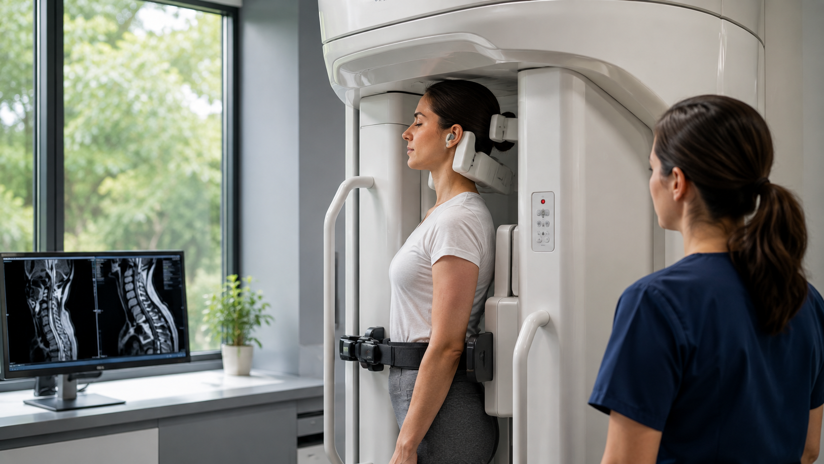
When a patient moves from supine to upright, the cervical spine experiences a fundamental change in the forces acting on it. In supine imaging, gravitational load on the cervical spine is essentially eliminated. The head is supported by the scanner table, the paraspinal muscles are relaxed, and the intervertebral discs are decompressed. The spine assumes a configuration that is determined primarily by the intrinsic resting tension of its passive structures rather than by the dynamic demands of supporting the head.
In the upright position, gravity acts continuously through the axial load of the head. The discs compress, the facet joints bear weight, and the posterior ligamentous structures are tensioned differently depending on the degree of curve the neck holds. In patients with cervical lordosis within normal limits, these changes are modest. In patients with flattened or reversed cervical curve, kyphotic deformity, or instability at specific segments, the changes under load are substantially greater and clinically meaningful.
The question of upright cervical MRI accuracy is most clearly answered in these load-sensitive cases: the upright study is not universally more accurate but is specifically more accurate for pathology that changes with loading and position, which is precisely the pathology most likely to be missed on a supine study.
Cervical Instability: A Case for Upright Imaging
Cervical instability, whether from congenital laxity, acquired ligamentous damage after trauma, inflammatory arthropathy, or connective tissue disorders, is a diagnosis that requires demonstrating abnormal movement between vertebral segments. This movement can only be captured when the spine is moving or under load. A supine neutral MRI can show the structural predisposing conditions but cannot measure the dynamic translation that constitutes instability.
Patients with suspected craniocervical instability, who may have conditions including Ehlers-Danlos syndrome, prior whiplash injury, or inflammatory arthropathy affecting the upper cervical joints, are among those who benefit most from upright positional cervical imaging. Measurements of the craniocervical junction in the upright position can reveal abnormal descent or distortion that is absent in the unloaded supine state. This is a finding that can change the clinical management entirely, from conservative management to neurosurgical evaluation.
The pattern of positional MRI hidden findings in the cervical region is particularly important because the consequences of missed cervical cord compression are more serious than missed compression at lumbar levels, where the cord itself is not at risk.
Myelopathy: When Cord Compression Is Position-Dependent
Cervical myelopathy, the syndrome of spinal cord dysfunction resulting from compression of the cord in the cervical canal, can present with a confusing picture when the compression is positional. A patient with myelopathic signs on examination who has a normal or near-normal supine MRI is one of the most important candidates for upright cervical imaging. The cord is being compressed in the upright or flexed position, producing the clinical signs, but the supine MRI captures the cord in its least compressed state.
This presentation is not rare. Patients with myelopathy secondary to dynamic cord compression from a combination of disc herniation in flexion and ligamentum flavum infolding in extension, or from positional craniocervical pathology, can have supine images that appear to show adequate canal dimensions while the cord is actually experiencing significant dynamic compression during daily activities. Imaging in the symptom-provoking position is the only way to capture this.
The evidence that traditional MRI missing pathology is particularly well-documented for dynamic cervical cord compression, where the gap between clinical findings and supine imaging results has prompted specialists to increasingly request positional studies when the clinical picture exceeds what the MRI shows.
Post-Traumatic Cervical Assessment
After cervical trauma, including motor vehicle accidents, contact sport injuries, and falls with head involvement, the ligamentous structures of the cervical spine may be damaged in ways that produce instability without obvious fracture or disc disruption on standard imaging.
The structural damage is to the ligaments themselves, which are not as visible on MRI as bony or disc pathology, and the consequence of that damage is dynamic: the spine moves abnormally under load and in specific positions.
Upright cervical MRI in flexion and extension after trauma provides the functional assessment of the cervical spine under the conditions that will govern the patient's recovery and their capacity to return to normal activities.
A patient cleared based on a supine MRI who has significant ligamentous instability in flexion may be cleared prematurely. The clinical and legal implications of missed post-traumatic cervical instability make upright imaging a particularly important consideration in this population.
The role of flexion and extension imaging in post-traumatic cervical assessment is recognised in clinical guidelines precisely because the functional consequences of missed instability can be severe and because supine imaging cannot adequately screen for it.
Patients With Symptoms Worse in Specific Neck Positions
A patient who reports that their symptoms are consistently better lying down and worse when upright, or who has pain or neurological symptoms that are provoked by specific head positions such as looking up, looking down, or rotating to one side, is describing a positional problem. The clinical pattern is the guide to the imaging approach: when symptoms are positional, the imaging should be performed in the symptomatic position.
Headaches that worsen when upright and improve with recumbency are a classic presentation associated with craniocervical pathology or cerebrospinal fluid dynamics that change with posture. Arm symptoms that are provoked by specific neck positions suggest a foraminal or dynamic compressive process rather than a fixed structural lesion. Both patterns indicate that the upright or positional scan is more likely to show the relevant pathology than the supine study.
For patients who have undergone standard imaging that did not explain their symptoms, upright MRI for chronic pain has provided the missing structural explanation in a significant proportion of cases, particularly when the symptoms were clearly position-dependent and had been attributed to non-structural causes.
Claustrophobia and Patient Comfort
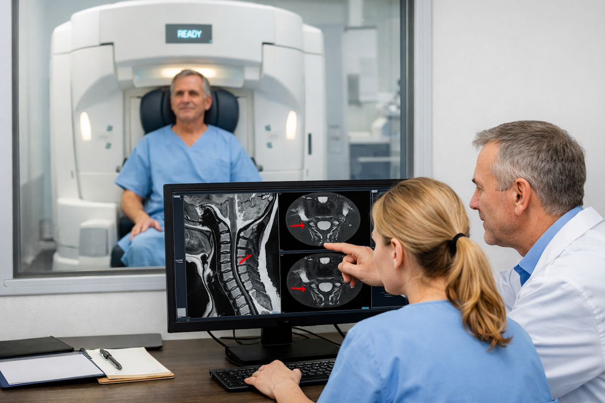
One practical advantage of upright open MRI that applies to cervical imaging specifically is the comfort and accessibility of the scanner design. Standard closed-bore cervical MRI requires the patient to lie supine with the head in a coil that surrounds the skull, inside a narrow tube.
For patients with claustrophobia, large body habitus, or neck pain that makes lying still in a constrained position very difficult, the supine study can be poorly tolerated or technically compromised by patient movement.
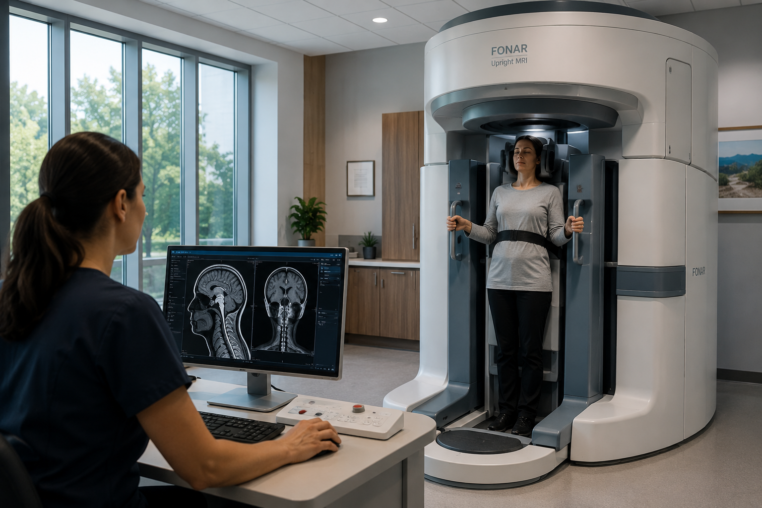
An upright open scanner performs the cervical study with the patient seated and unrestricted by an enclosing bore. The exam can be performed with better patient comfort, which often translates to less motion artifact and better image quality in patients who struggle with conventional scanners.
For cervical imaging specifically, where subtle findings related to cord compression and foraminal dimensions require high image quality, patient cooperation is a meaningful factor in study quality.
Information about the spine MRI services at Upright MRI of Deerfield, including cervical protocols and how to request positional sequences, is available for patients and referring clinicians considering whether an upright study is appropriate.
Frequently Asked Questions
Is upright cervical MRI image quality as good as a high-field closed scanner?
Upright open MRI systems typically operate at lower field strengths than the highest-field closed scanners, which affects signal-to-noise ratio and fine resolution. For the specific clinical questions where upright imaging adds value, including instability, dynamic compression, and positional findings, the diagnostic benefit of imaging in the relevant position outweighs the technical resolution advantage of higher-field supine imaging. For specific soft tissue detail questions, closed scanners may retain an advantage.
What neck conditions benefit most from upright MRI?
Cervical instability of any cause including connective tissue disorders, post-traumatic ligamentous injury, and inflammatory arthropathy; position-dependent myelopathy; dynamic foraminal stenosis; symptoms provoked by specific head positions; and any presentation where the supine MRI findings do not adequately explain the clinical picture.
Can the flexion and extension positions be combined with upright cervical imaging?
Yes. In an upright positional scanner, the cervical spine can be imaged in neutral upright, maximum flexion, and maximum extension in a single session. The comparative measurements across these positions provide the full dynamic picture of segmental movement and canal dimensions at each position.
How do I describe my symptoms to help determine if upright cervical MRI is right for me?
Note specifically which positions or activities make your symptoms better or worse. If lying down relieves your symptoms and sitting or standing worsens them, that is important positional information. If looking up, looking down, or rotating to one side provokes arm pain, tingling, or weakness, record that specifically. The more precisely you can describe the positional pattern, the more accurately the imaging can be targeted.
Will a referring physician need to specifically request the upright or positional sequences?
Yes. A standard cervical spine MRI referral will typically be performed in the scanner's default neutral supine or neutral seated position. To obtain flexion and extension sequences, or to specify that the exam should be performed in a position that reproduces symptoms, this needs to be explicitly stated in the referral or discussed with the imaging centre before the appointment.
The Bottom Line
A neck MRI is better performed in an upright scanner when symptoms are position-dependent, when instability is suspected, when myelopathic signs exceed what the supine MRI shows, when post-traumatic ligamentous injury needs functional assessment, or when patient factors make supine scanning difficult. The upright scanner captures the cervical spine under the gravitational and positional conditions that govern the patient's symptoms, which is the clinically relevant information for diagnosis and treatment planning.
Upright MRI of Deerfield performs cervical spine imaging in upright, flexion, and extension positions for patients referred from across the Chicago area and Midwest. If a supine neck MRI has not explained your symptoms, or if your clinician wants a positional study, contact the team to discuss the right protocol for your situation.
SHARE THIS POST:
Leave a Comment:

The World's Most Patient-Friendly MRI. A comfortable, stress-free, and completely reliable MRI scan. We offer patients an open, upright, standup MRI experience that helps those who are claustrophobic and stress being in a confined area. Upright MRI of Deerfield is recognized as the world leader in open MRI innovation,
Our Recent Post


READ PATIENT TESTIMONIALS
Upright MRI of Deerfield.
Susan D.,
Highland Park, 39
I am going to tell everyone about your office! This was a great experience after I panicked in other MRI machines and had to leave. Thank you so much.

Judith B.,
Milwaukee, 61
I suffer from vertigo and other MRIs do not work. This was wonderful…absolutely NO discomfort at all. The MRI was so fast…I wanted to stay and watch the movie! Mumtaz was great. His humor really put me at ease. I’ve already recommended Upright MRI to friends.
Delores P.,
Glencoe, 55
Everything is so nice and professional with your place. I have been there a couple of times. My husband and I would not go anywhere else.