
How Does an Upright Brain MRI Improve CSF Analysis?
Cerebrospinal fluid is one of the most diagnostically important substances in the body, and one of the most underappreciated. This clear fluid surrounds and cushions the brain and spinal cord, provides nutrients to neural tissue, removes waste products, and regulates pressure within the skull and spinal canal. When CSF flow or pressure is abnormal, the consequences can be serious and wide-ranging. But capturing what is actually happening with CSF in a clinically meaningful way is harder than it sounds.
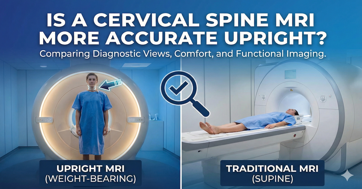
The challenge is that CSF dynamics change significantly depending on body position. When a person is lying flat, gravity no longer acts on the brain and cerebellar structures the way it does when they are upright. The distribution of intracranial pressure, the position of the cerebellar tonsils, and the flow characteristics of CSF through key channels all change in response to gravitational load. A scan taken lying down captures the system at rest in an artificial position. An upright scan captures it under the conditions the patient actually lives in.
Quick Answer: An upright brain MRI improves CSF analysis by imaging the brain and its surrounding fluid under the effects of gravity, which is the position in which symptoms typically occur. This produces a more accurate picture of CSF flow, intracranial pressure distribution, and structural relationships that affect cerebrospinal fluid dynamics. For conditions like Chiari malformation, cranio-cervical instability, and intracranial hypertension, upright imaging frequently reveals abnormalities that lying-down scans do not show.

What CSF Does and Why Its Dynamics Matter
Cerebrospinal fluid circulates through the brain's ventricles and around the spinal cord in a continuous, pulsatile flow synchronized with the heartbeat. It is produced mainly in the choroid plexus within the ventricles and absorbed into the venous system. The total volume of CSF in an adult is modest, around 150 milliliters, but its pressure and flow characteristics have a significant influence on neurological function.
When CSF flow is obstructed or when the balance between production and absorption is disrupted, pressure builds. When structures shift in ways that compress the pathways through which CSF moves, flow patterns change. Both scenarios can produce symptoms that range from headaches that worsen with position change, to cognitive difficulties, vision changes, neck pain, and a constellation of neurological symptoms that are sometimes attributed to multiple unrelated causes before the underlying CSF abnormality is identified.
The brainstem junction where the brain meets the cervical spine is a particularly important region for CSF dynamics. The foramen magnum, the opening at the base of the skull, is the primary channel through which CSF passes between the cranial and spinal compartments. Any structural issue that affects the size or function of this channel, whether from a tonsillar herniation, instability, or another cause, can directly affect how CSF moves.
Why Position Changes What the Scan Shows
The position of the patient during imaging is not incidental. Why lying-down imaging misses critical findings applies as directly to brain and CSF assessment as it does to spinal diagnosis: the supine position removes the gravitational load that shapes how structures are arranged and how CSF moves when the patient is actually experiencing symptoms.
In a lying-down scan, the cerebellar tonsils may not herniate to the degree they do when the patient is upright. Intracranial pressure distribution changes. Venous drainage patterns shift. The brainstem can reposition relative to the surrounding anatomy. For patients whose symptoms are clearly worse when upright and better when lying down, this positional difference is the entire diagnostic story. A scan that captures only the lying-down state is, in those cases, capturing the state in which the patient has fewest symptoms.
Upright brain MRI captures the anatomy under gravitational load. Structures are in the positions they occupy during daily life. CSF pathways are under the pressures they experience when the patient is standing or sitting. The scan shows what is actually happening when the patient feels worst, which is the information most relevant to diagnosis and treatment planning.
Chiari Malformation and Positional CSF Obstruction
Chiari malformation, in which the cerebellar tonsils descend through the foramen magnum and compress the upper cervical spinal canal, is one of the conditions where upright brain imaging produces the most clinically significant improvements in CSF analysis. Chiari malformation diagnosis depends on position in a meaningful way: the degree of tonsillar herniation can be measurably different between lying and upright positions, and the obstruction of CSF flow at the foramen magnum may only be visible in the upright state.
Patients with Chiari who have symptoms that worsen when upright and improve when lying down are describing, in clinical terms, exactly the positional CSF obstruction that an upright scan is designed to capture. When a lying-down scan shows borderline or minimal tonsillar descent in these patients, the clinical picture is incomplete. The upright scan provides the missing dimension.
Beyond the structural component, CSF flow imaging studies performed in the upright position can document the actual obstruction of fluid movement at the foramen magnum in ways that lying-down studies may not. This information is directly relevant to treatment decisions, including whether surgical intervention is appropriate and what approach is most likely to restore normal CSF dynamics.
Cranio-Cervical Instability and Its Effect on CSF
Cranio-cervical instability, in which the ligamentous structures that stabilize the junction between the skull and the upper cervical spine are compromised, affects CSF dynamics in several ways. Abnormal movement at this junction can create mechanical effects on the brainstem and upper cervical spinal cord that intermittently compress CSF pathways. Cranio-cervical instability imaging requires capturing the anatomy under dynamic conditions, including the positional stresses that provoke symptoms.
The challenge with CCI diagnosis in general is that conventional lying-down imaging shows the structures at rest, when the instability may not be manifest. Positional MRI reveals CCI by capturing the anatomy in positions that load the craniocervical junction and allow abnormal movement to become visible on imaging.
The connection to CSF analysis is that when the craniocervical junction is unstable, CSF pathways can be intermittently compressed in ways that a resting scan will not show. Patients with CCI often describe symptoms that are highly position-dependent, including pressure symptoms in the head, cognitive symptoms, and sensory changes that correlate with specific head positions or activities. Understanding this connection between structural instability and CSF dynamics often requires the positional context that only an upright scan provides.
Intracranial Hypertension and Positional Pressure Changes
Intracranial hypertension, whether idiopathic or secondary to another cause, involves abnormally elevated pressure within the skull. This pressure is not constant and can change significantly with position. Many patients with intracranial hypertension describe symptoms that are notably worse when lying down and somewhat better when upright, because gravity assists venous drainage from the cranial compartment in the upright position.
For other patients, symptoms are worse upright, which can reflect a different pattern of pressure dynamics. Understanding which positional pattern applies to a given patient has diagnostic and treatment implications. Upright brain imaging provides structural context that helps interpret the pressure patterns and the anatomical factors that may be contributing to them.
The brain MRI scan at Upright MRI of Deerfield provides the positional imaging context that is essential for this kind of assessment, capturing the brain and its surrounding structures under the gravitational conditions that most closely match daily life.
Other Conditions Where Upright Brain CSF Analysis Adds Value
Beyond Chiari and CCI, there are several conditions where upright brain imaging improves the quality of CSF-related assessment. Conditions upright MRI detects that standard scans miss include syringomyelia, where fluid-filled cavities form within the spinal cord secondary to impaired CSF dynamics, and some forms of hydrocephalus where the pattern of ventricular enlargement and CSF distribution may look different under gravitational load.
Patients who have experienced traumatic brain injury may also benefit from upright brain imaging. Upright MRI for concussions and brain injuries can reveal structural changes and CSF flow abnormalities that contribute to persistent post-concussion symptoms, particularly those that vary with position.
For patients with multiple sclerosis, where intracranial pressure and CSF dynamics can be affected by the disease process, upright MRI and multiple sclerosis diagnosis represents an area of growing clinical interest.
What the Scan Experience Looks Like
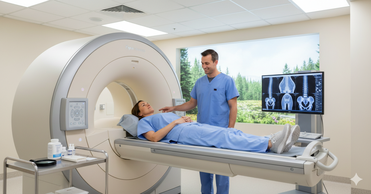
An upright brain MRI is performed with the patient seated or standing within an open magnet configuration rather than lying inside a closed tunnel. The patient's head is positioned in the imaging field while they are in a natural, gravitationally loaded position. The scan captures the brain, posterior fossa, brainstem, and upper cervical structures in this position, as well as in any additional positions the referring physician specifies.
The open configuration means there is no enclosed tunnel and significantly less claustrophobic experience than a conventional MRI. For patients with conditions that make lying still in a closed environment difficult, or for those who need flexion and extension images of the cervical spine as part of the same assessment, the upright format provides both clinical and practical advantages over conventional scanning.
Frequently Asked Questions
Can an upright MRI measure actual CSF pressure?
Standard MRI, whether upright or lying down, provides structural and flow imaging rather than direct pressure measurement. CSF pressure is typically measured through lumbar puncture. However, upright MRI can identify structural findings and flow patterns that are strongly associated with elevated or abnormal pressure, providing valuable supporting information for clinical interpretation.
Is upright brain MRI appropriate for all patients with Chiari malformation?
Upright brain MRI is particularly valuable for patients with borderline Chiari findings on lying-down scans who have clear positional symptoms, and for those being evaluated for surgical intervention. The imaging provides positional context that can significantly change the clinical picture. Your physician can advise whether it is appropriate for your specific situation.
How does upright brain MRI differ from a standard brain MRI in terms of what it shows?
Both types of scan use the same magnetic resonance imaging principles and can show the same brain structures. The key difference is that upright brain MRI shows those structures under gravitational load, which changes the position of the cerebellum, the tension on posterior fossa structures, and the distribution of intracranial pressure in ways that are diagnostically relevant for position-sensitive conditions.
What symptoms might indicate that a positional brain MRI is appropriate?
Headaches that worsen significantly with standing or sitting and improve when lying down, symptoms that are clearly worse with specific head positions, pressure or fullness at the base of the skull, vision changes associated with position, and cognitive symptoms that fluctuate with position are all patterns that suggest positional imaging would add valuable information.
Does an upright brain MRI require a referral?
A referral from a physician is recommended and ensures the imaging is interpreted in the context of your clinical history. Many patients come to Upright MRI of Deerfield having already had a conventional brain MRI, and their physician wants the additional positional information to complete the diagnostic picture. Consulting with the facility directly can clarify the specific requirements for your situation.
The Bottom Line
Cerebrospinal fluid dynamics are position-dependent, and capturing them under gravitational load provides diagnostic information that lying-down brain scans cannot offer for position-sensitive conditions. For patients with Chiari malformation, cranio-cervical instability, intracranial hypertension, and related conditions, upright brain MRI is not a marginal upgrade but a meaningfully different diagnostic tool.
Upright MRI of Deerfield performs brain and cranio-cervical imaging in the upright position that provides the positional context essential for these assessments. If you or your physician believe positional brain imaging could add important information to your diagnostic picture, reaching out to discuss your specific situation is the right next step.
SHARE THIS POST:
Leave a Comment:

The World's Most Patient-Friendly MRI. A comfortable, stress-free, and completely reliable MRI scan. We offer patients an open, upright, standup MRI experience that helps those who are claustrophobic and stress being in a confined area. Upright MRI of Deerfield is recognized as the world leader in open MRI innovation,
Our Recent Post


READ PATIENT TESTIMONIALS
Upright MRI of Deerfield.
Susan D.,
Highland Park, 39
I am going to tell everyone about your office! This was a great experience after I panicked in other MRI machines and had to leave. Thank you so much.

Judith B.,
Milwaukee, 61
I suffer from vertigo and other MRIs do not work. This was wonderful…absolutely NO discomfort at all. The MRI was so fast…I wanted to stay and watch the movie! Mumtaz was great. His humor really put me at ease. I’ve already recommended Upright MRI to friends.
Delores P.,
Glencoe, 55
Everything is so nice and professional with your place. I have been there a couple of times. My husband and I would not go anywhere else.

