Why Is an Upright MRI Essential for Diagnosing Chiari Malformation?
Chiari malformation is a neurological condition that can lead to serious symptoms like headaches, neck pain, and dizziness. However, diagnosing Chiari malformation is not always straightforward, especially since its symptoms can be easily mistaken for other conditions. Traditional imaging methods, such as X-rays or MRIs, sometimes fail to fully capture the complexities of the malformation. This is where Upright MRI technology comes into play. It has become an essential tool for diagnosing Chiari malformation, offering clear advantages over traditional MRI techniques by providing dynamic, functional images that help doctors better understand the condition’s impact.
What Is Chiari Malformation?
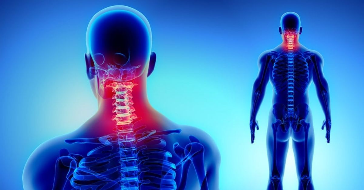
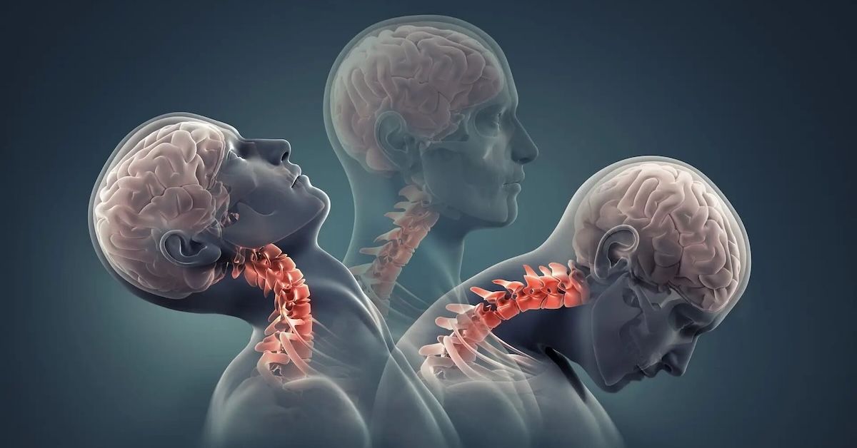
Chiari malformation occurs when part of the brain, specifically the cerebellum, extends into the spinal canal. This can cause a range of symptoms depending on the type of Chiari malformation (Type I or Type II), and the severity of the herniation. Common symptoms include severe headaches, neck pain, dizziness, and difficulty with balance and coordination. In some cases, patients may also experience numbness or vision problems.
Diagnosing Chiari malformation can be challenging, especially since its symptoms can overlap with other conditions like migraines or vertigo. Traditional diagnostic tools like X-rays often fail to reveal the full extent of the condition, particularly in cases where the herniation is not severe or the symptoms fluctuate depending on posture.
Traditional MRI and Its Limitations
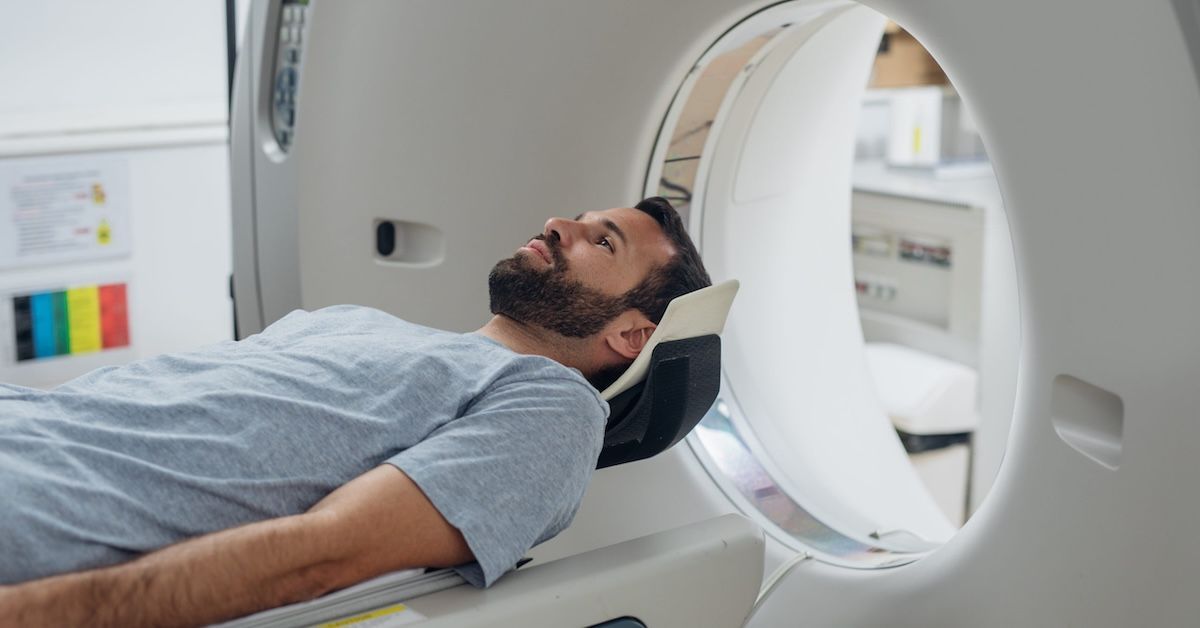
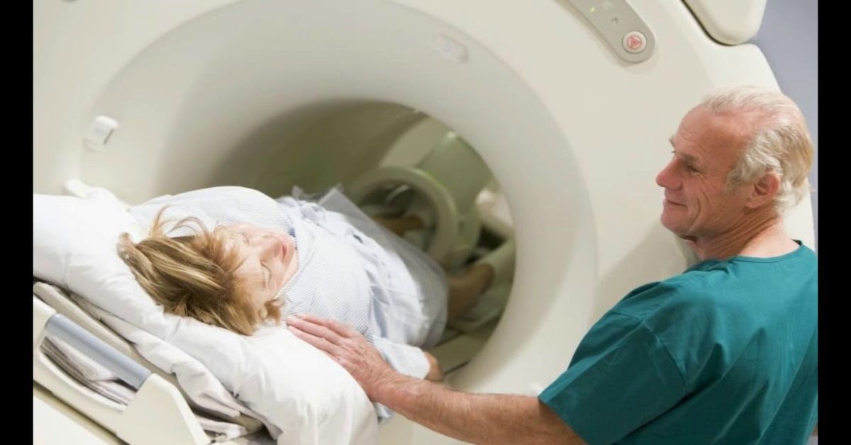
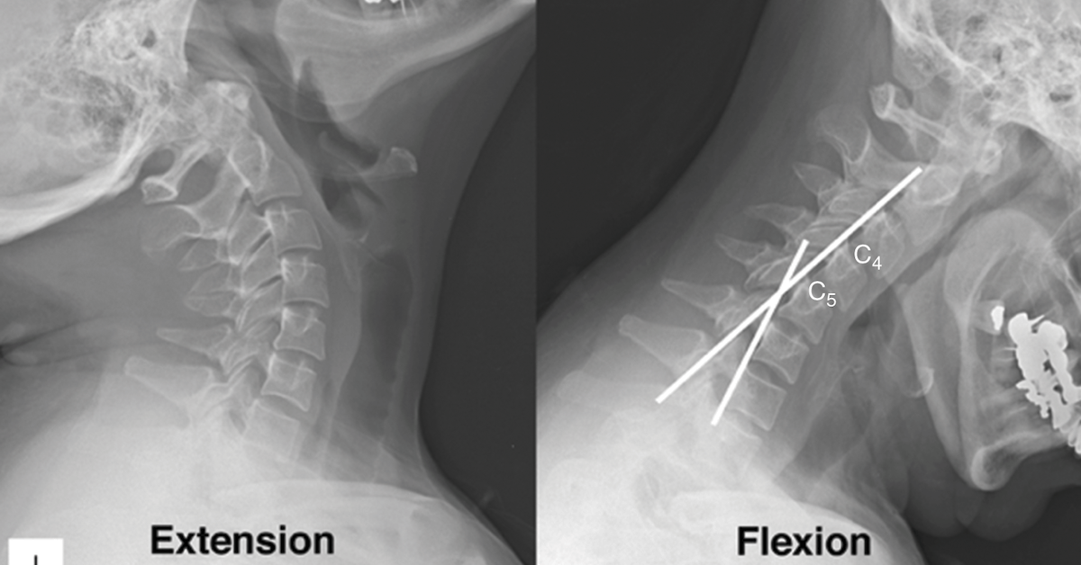
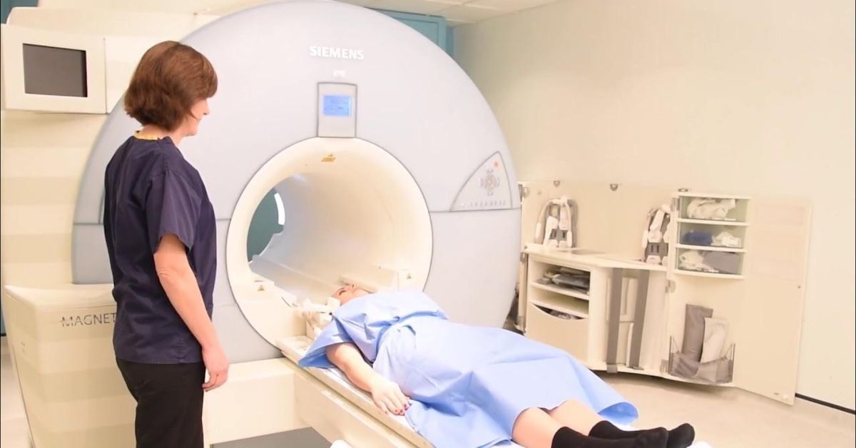
Traditional MRI technology provides clear images of the brain and spinal cord. However, these images are static and show the body in a non-weight-bearing, prone position. For conditions like Chiari malformation, where symptoms are often aggravated by changes in posture or gravity, traditional MRIs may miss key signs of the condition.
In a typical MRI scan, the patient is required to lie flat, which does not replicate the physical stresses that the body experiences in an upright position. For Chiari malformation, where the herniation of the cerebellum is often aggravated by the effects of gravity, lying flat on the table may not produce the same symptoms or reveal the same abnormalities that occur when the patient is standing or sitting.
What Is an Upright MRI?
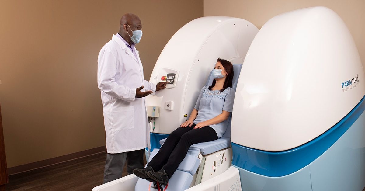
Upright MRI technology allows patients to be scanned in an upright position, either standing or seated, which is a significant departure from traditional MRIs that require the patient to lie flat. This unique positioning allows for dynamic imaging of the brain and spine, better replicating the way these structures interact during daily activities, such as standing, walking, or moving.
By capturing images in a more natural, functional position, Upright MRI provides a much more realistic view of the brain and spine under the effects of gravity. For patients with Chiari malformation, this means that the herniation of the cerebellum and the impact of the malformation on the spinal cord can be captured while the patient is in a position that mimics real-life conditions.
How Upright MRI Enhances the Detection of Chiari Malformation
One of the main reasons Upright MRI is crucial for diagnosing Chiari malformation is its ability to show how the condition responds to the forces of gravity. In many cases, Chiari malformation symptoms worsen when the patient is upright, making it essential to capture this dynamic aspect of the condition during the imaging process. Traditional MRI simply cannot replicate these movements and forces.
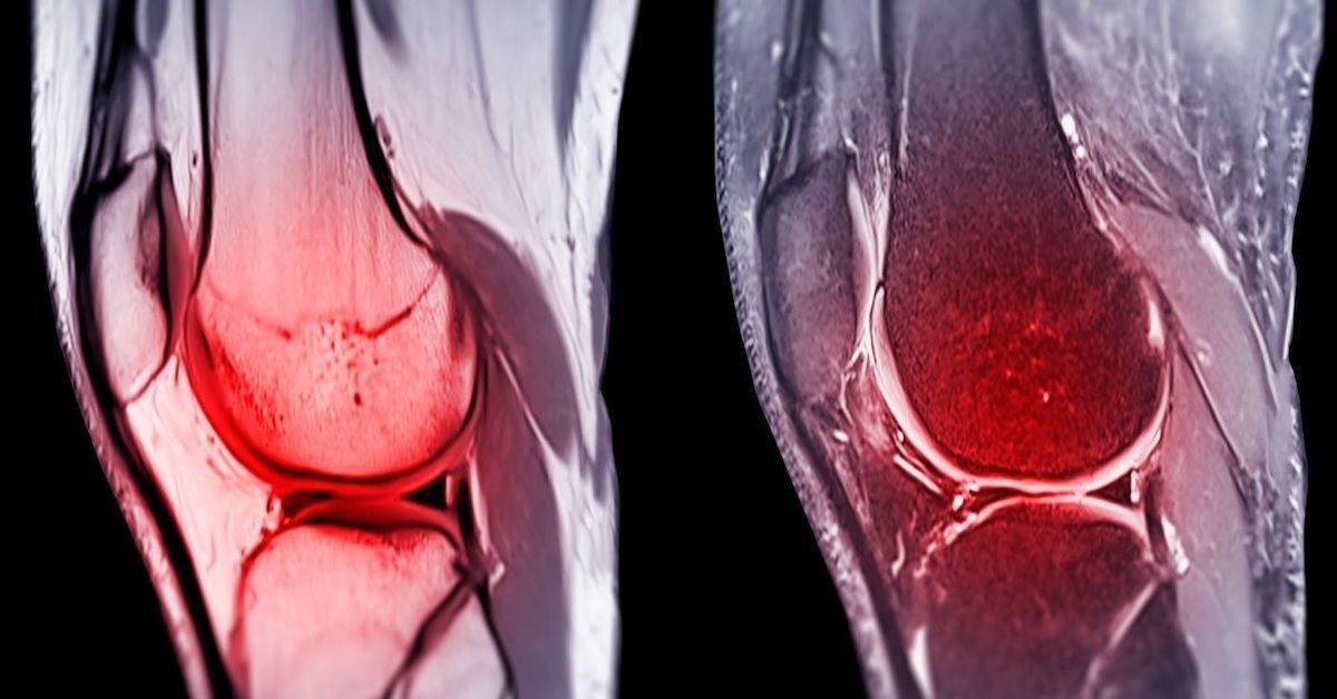
By using Upright MRI, healthcare providers can observe how the brain and spinal cord interact in real-world conditions. This allows for better visualization of the brain tissue that is herniated and may be causing compression on the spinal cord or other structures. It also helps reveal any subtle changes in the position of the cerebellar tonsils, which may not be immediately noticeable in a traditional lying-down MRI scan.
Furthermore, Upright MRI provides a clearer picture of spinal cord compression, which is common in Chiari malformation cases. As gravity plays a significant role in worsening the symptoms, an Upright MRI captures this phenomenon, providing doctors with valuable insights that may not have been visible in a traditional scan.
Why Upright MRI Is Essential for Chiari Malformation Diagnosis
The ability to capture the effects of gravity and real-world movement makes Upright MRI a game-changer in diagnosing Chiari malformation. Not only does it offer more accurate, functional imaging of the brain and spine, but it also allows doctors to see how the malformation behaves in different postures. This gives healthcare providers a more complete understanding of the severity and progression of the condition, leading to better-informed treatment decisions.
Upright MRI allows for clearer images of the spinal cord, brain, and surrounding tissues. It helps identify not only Chiari malformation but also any secondary conditions that may have developed, such as syringomyelia (a cyst or cavity within the spinal cord), which is often associated with Chiari malformation. By providing a more comprehensive view of the condition, Upright MRI plays a critical role in formulating a targeted treatment plan.
Additional Benefits of Upright MRI for Chiari Malformation
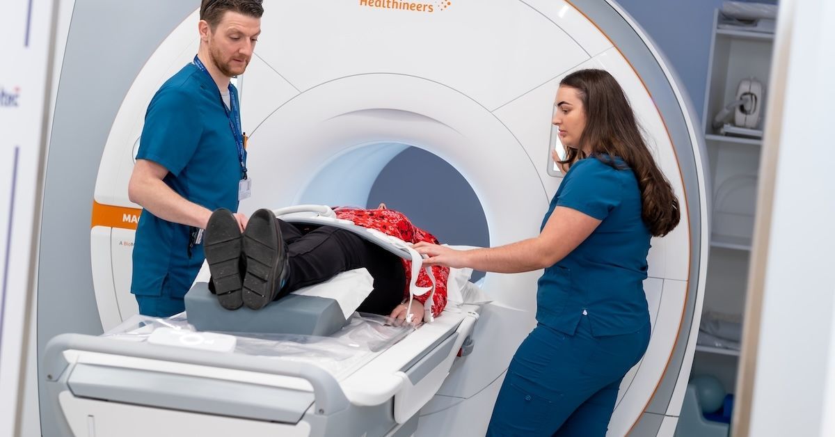
Beyond improving diagnostic accuracy, Upright MRI offers several other advantages for patients with Chiari malformation. First, it provides a more comfortable and patient-friendly experience. Many individuals with Chiari malformation experience neck pain or discomfort, which can make lying flat for an extended period difficult. The ability to undergo imaging in an upright position reduces the discomfort and makes the entire process more tolerable.
Upright MRI also reduces the need for multiple diagnostic tests. Traditional MRI scans may not provide a clear diagnosis, leading to additional imaging or tests. With Upright MRI, healthcare providers can gather more comprehensive data in one session, saving time and reducing the number of follow-up appointments needed.
Conclusion
In conclusion, Upright MRI is an essential tool for accurately diagnosing Chiari malformation. Unlike traditional MRI, which provides static images that may miss key aspects of the condition, Upright MRI captures the dynamic effects of gravity and movement, offering a more realistic view of how the malformation affects the brain and spine. This allows healthcare providers to make more accurate diagnoses and develop more effective treatment plans.
If you are experiencing symptoms of Chiari malformation or suspect you have this condition, an Upright MRI could provide the answers you need. At Upright MRI of Deerfield, we offer state-of-the-art Upright MRI technology to help you get the most accurate diagnosis possible. Contact us today to learn more about how Upright MRI can help you on your path to recovery.
SHARE THIS POST:
Leave a Comment:

The World's Most Patient-Friendly MRI. A comfortable, stress-free, and completely reliable MRI scan. We offer patients an open, upright, standup MRI experience that helps those who are claustrophobic and stress being in a confined area. Upright MRI of Deerfield is recognized as the world leader in open MRI innovation,
Our Recent Post

READ PATIENT TESTIMONIALS
Upright MRI of Deerfield.
Susan D.,
Highland Park, 39
I am going to tell everyone about your office! This was a great experience after I panicked in other MRI machines and had to leave. Thank you so much.

Judith B.,
Milwaukee, 61
I suffer from vertigo and other MRIs do not work. This was wonderful…absolutely NO discomfort at all. The MRI was so fast…I wanted to stay and watch the movie! Mumtaz was great. His humor really put me at ease. I’ve already recommended Upright MRI to friends.
Delores P.,
Glencoe, 55
Everything is so nice and professional with your place. I have been there a couple of times. My husband and I would not go anywhere else.


