
Decoding the Hidden Secrets: MRI Knee Scans Unveiled
The human knee is a complex joint that is necessary for daily activities and movement. However, it is prone to a number of illnesses and injuries that can impair its functionality and be uncomfortable, just like any other part of our body. Here, magnetic resonance imaging (MRI) is useful because it provides a window into the knee joint's well-kept secrets. We will dig into the interesting world of MRI knee scans in this extensive analysis, from the technology underlying them to the plethora of diseases they may reveal.
Beyond the Surface: MRI Magic
Strong magnetic fields and radio waves are used in the extraordinary medical imaging method known as magnetic resonance imaging, or MRI, to provide precise pictures of the inside organs of the body. Due to its special capacity to deliver high-resolution pictures of the knee ligaments, tendons, cartilage, and bones, MRI takes center stage when it comes to knee scans. MRI uses no ionizing radiation, unlike X-rays or CT scans, making it a safe and popular option for knee examinations.
Common Knee Conditions Diagnosed by MRI
Ligament Tears: The Crucial Connectors
The medial collateral ligament (MCL), lateral collateral ligament (LCL), posterior cruciate ligament (PCL), and anterior cruciate ligament (ACL) all play important roles in preserving knee stability. Knee function may be significantly impacted by injuries to these ligaments. MRI is helpful in identifying and assessing ligament tears, which enables doctors to choose the best course of therapy, whether that be conservative methods or surgical surgery.
Meniscal Tears: Protecting the Cushion
A C-shaped piece of cartilage in the knee called the meniscus serves as a cushion and stabilizes the joint. Meniscal rips are frequent and can be caused by a variety of traumas or activities. For orthopedic experts to create individualized treatment programs, which may involve rest, physical therapy, or arthroscopic surgery, it is essential that MRI be able to see and diagnose meniscal tears.
Cartilage Damage: The Shock Absorbers
The knee's cartilage acts as a shock absorber, protecting the joint during motion. A kind of cartilage injury known as osteochondral defects can cause discomfort and restricted movement. When it comes to identifying cartilage damage and determining its degree, MRI is excellent. This helps medical professionals make judgments regarding treatment options like joint replacement surgery or cartilage repair methods.
Tendon Injuries: The Powerhouses
Strong connective structures called tendons hold muscles to bones, allowing for easier joint mobility. Overuse or repeated stress can lead to injuries like tendinitis and tendinosis. With the use of MRI, these disorders may be accurately diagnosed, allowing for more focused treatment plans that may include rest, physical therapy, or, in more serious circumstances, surgical repair.
Arthritis: The Joint Invaders
Osteoarthritis and rheumatoid arthritis are two types of arthritis that can damage the knee joint. Joint inflammation, cartilage deterioration, and bone alterations are caused by these disorders. Rheumatologists and orthopedic surgeons can develop effective treatment programs that may involve medication, physical therapy, or joint injections by using MRI to determine the amount of joint damage.
Fractures and Bone Bruises: The Silent Threats
Injuries such as fractures and bone bruises can significantly impact the knee joint's structural integrity. MRI is a valuable tool for detecting and classifying these injuries, enabling physicians to make timely decisions about immobilization, surgical intervention, or other necessary treatments.
Preparing for an MRI Knee Scan
The structural integrity of the knee joint can be dramatically impacted by wounds like fractures and bone bruising. MRI is a useful tool for identifying and diagnosing these injuries, giving doctors the information they need to decide when immobilization, surgical intervention, or other essential therapies are the best course of action.
The MRI Knee Scan Process
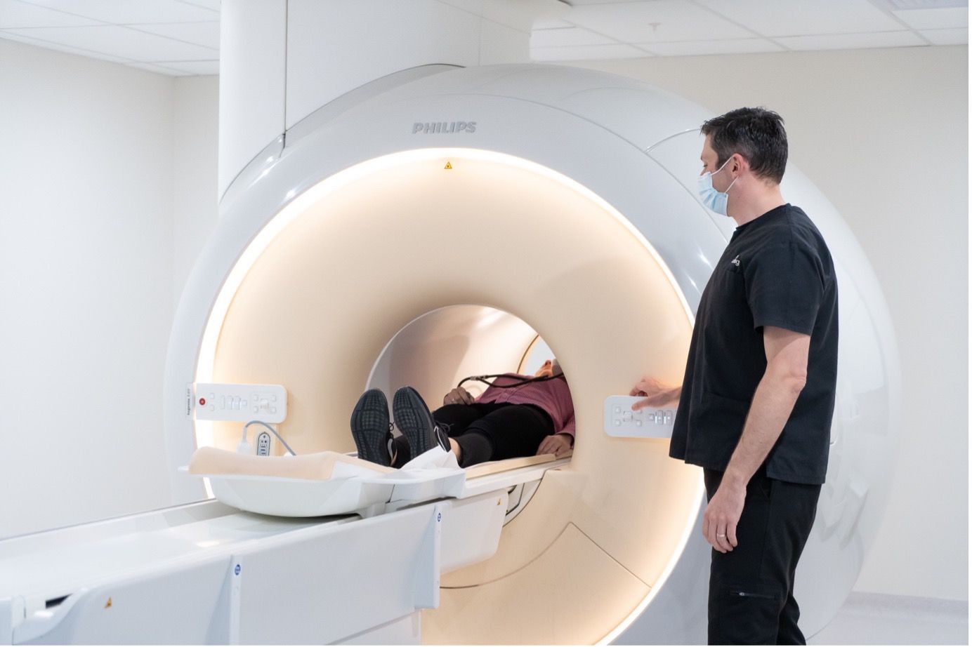
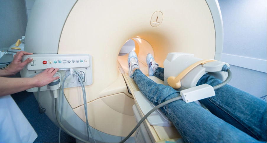
The MRI knee scan itself is a pretty quick and painless process. The afflicted knee is placed in a customized coil as the patient lies comfortably on an examination table. To achieve accurate and clear pictures throughout the scan, it's important to maintain as much stillness as possible. A radiologic technologist interacts with the patient during the procedure, giving instructions and assuring their comfort. The length of the scan might range from 30 minutes to more than an hour, depending on how complicated the examination is.
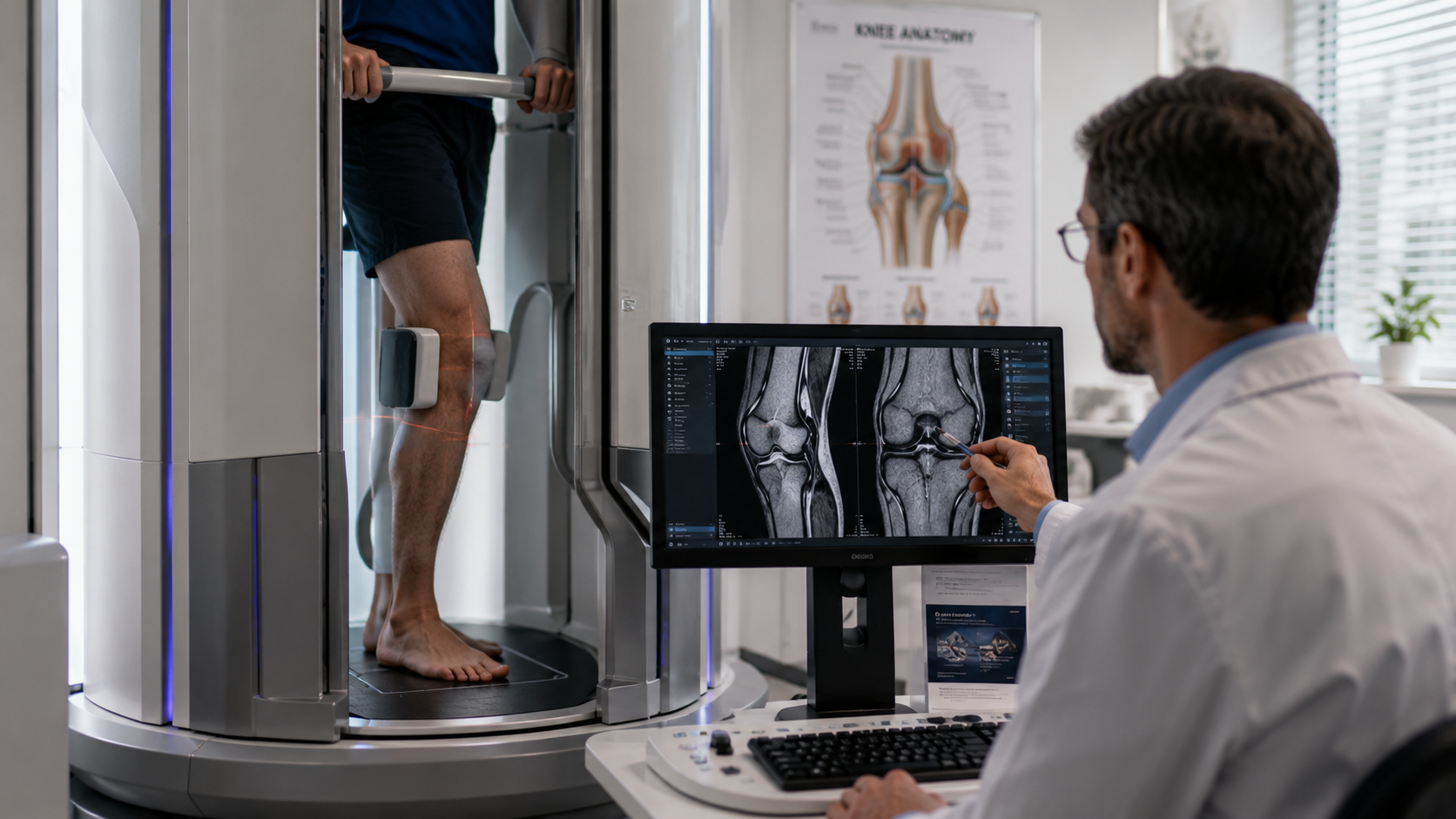
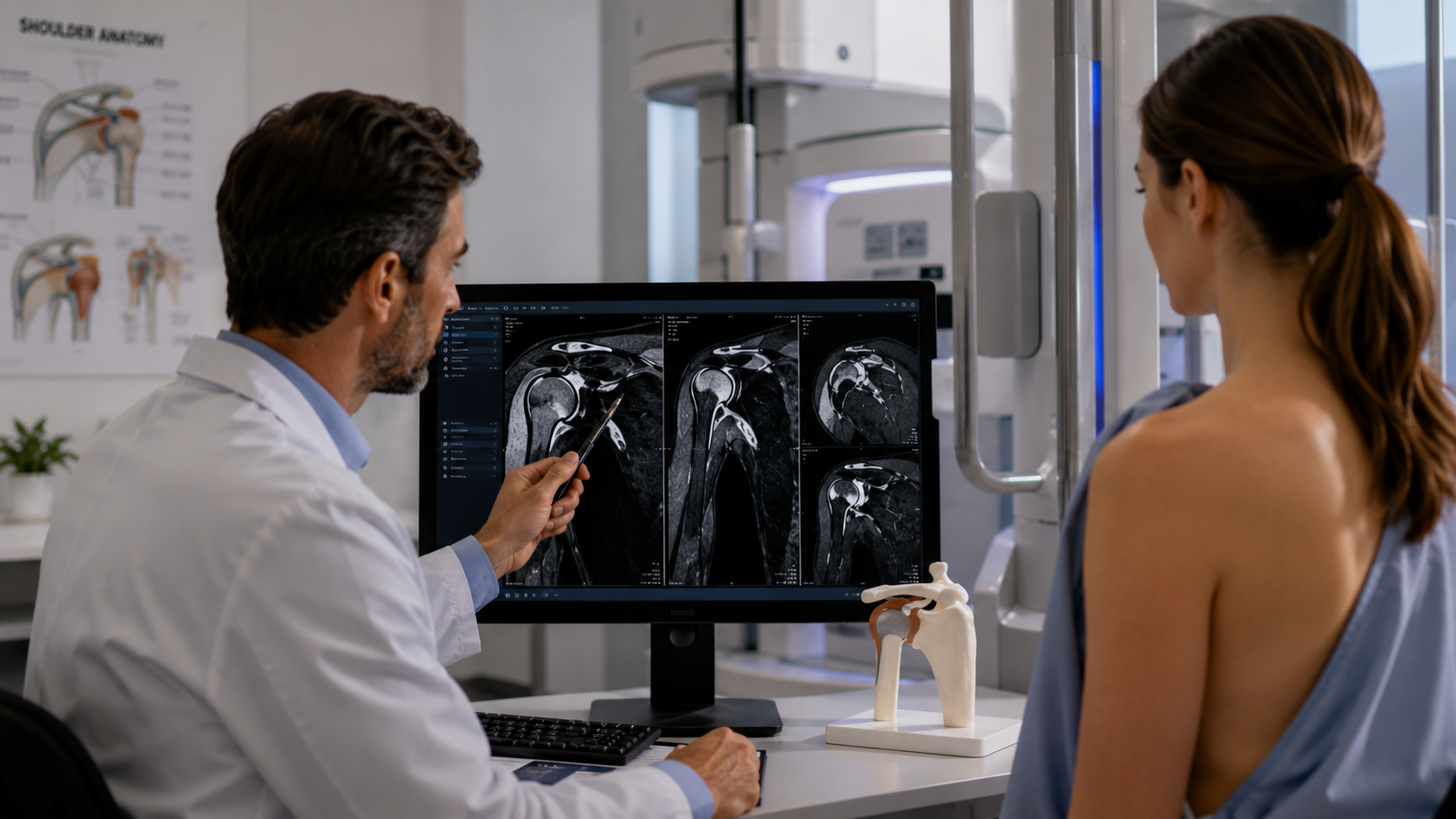
Interpreting MRI Knee Scan Results
Your knee's MRI pictures are taken, and then they go through a comprehensive review by trained radiologists. These specialists carefully examine the photos in search of particular clues about the condition they are evaluating. The results are then thoroughly documented in a report that offers your referring doctor useful information. Although some of the language used in these reports may sound technical, your healthcare professional will walk you through the findings and explain what they indicate for the health of your knees.
Benefits and Limitations of MRI for Knee Diagnoses
There are several benefits of using MRI to diagnose knee issues. It is a popular option since it can deliver high-resolution pictures of soft tissues without subjecting patients to ionizing radiation. However, there are some circumstances when other imaging techniques, including X-rays or CT scans, may support the diagnosis, particularly when determining the bone structure is necessary. The best course of action will be decided by your healthcare professional depending on your unique circumstances.
Future Trends in Knee Imaging
Both technology and the subject of knee imaging are constantly evolving. New approaches offer scan times that are shorter and photos with even greater quality. Furthermore, advancements in imaging techniques and contrast agents have the potential to substantially improve diagnostic precision. Keeping up with these changes can help patients and medical professionals make the best choices for their knee care.
Conclusion
The mysteries of knee health have been revealed by magnetic resonance imaging, which has made it possible to accurately and early diagnose a variety of problems. MRI knee scans offer the essential information required to direct your therapeutic journey, regardless of whether you are battling with ligament tears, cartilage damage, or any other knee disease. Never forget that Upright MRI of Deerfield and the rest of your healthcare team are here for you at every turn. Accept the power of information and take initiative to keep your knees healthy and pain-free.
SHARE THIS POST:
Leave a Comment:

The World's Most Patient-Friendly MRI. A comfortable, stress-free, and completely reliable MRI scan. We offer patients an open, upright, standup MRI experience that helps those who are claustrophobic and stress being in a confined area. Upright MRI of Deerfield is recognized as the world leader in open MRI innovation,
Our Recent Post


READ PATIENT TESTIMONIALS
Upright MRI of Deerfield.
Susan D.,
Highland Park, 39
I am going to tell everyone about your office! This was a great experience after I panicked in other MRI machines and had to leave. Thank you so much.

Judith B.,
Milwaukee, 61
I suffer from vertigo and other MRIs do not work. This was wonderful…absolutely NO discomfort at all. The MRI was so fast…I wanted to stay and watch the movie! Mumtaz was great. His humor really put me at ease. I’ve already recommended Upright MRI to friends.
Delores P.,
Glencoe, 55
Everything is so nice and professional with your place. I have been there a couple of times. My husband and I would not go anywhere else.

