What Should I Understand About MRI for Arm and Hand Issues?
If you’ve been struggling with arm or hand pain that just won’t go away, you may have heard your doctor mention getting an MRI. You might be wondering, “What exactly does an MRI show that other tests don’t?” And more importantly, “How can it help me find relief?”
Magnetic Resonance Imaging (MRI) is a powerful tool that gives doctors detailed images of what’s going on inside your body, especially when it comes to areas like the arm and hand where soft tissue injuries are common. This non-invasive, radiation-free test can help get to the bottom of pain, nerve issues, or other conditions that might be difficult to diagnose with traditional imaging like X-rays. Let’s dive into everything you need to know about MRI for arm and hand issues.
What Is MRI and How Does It Work?
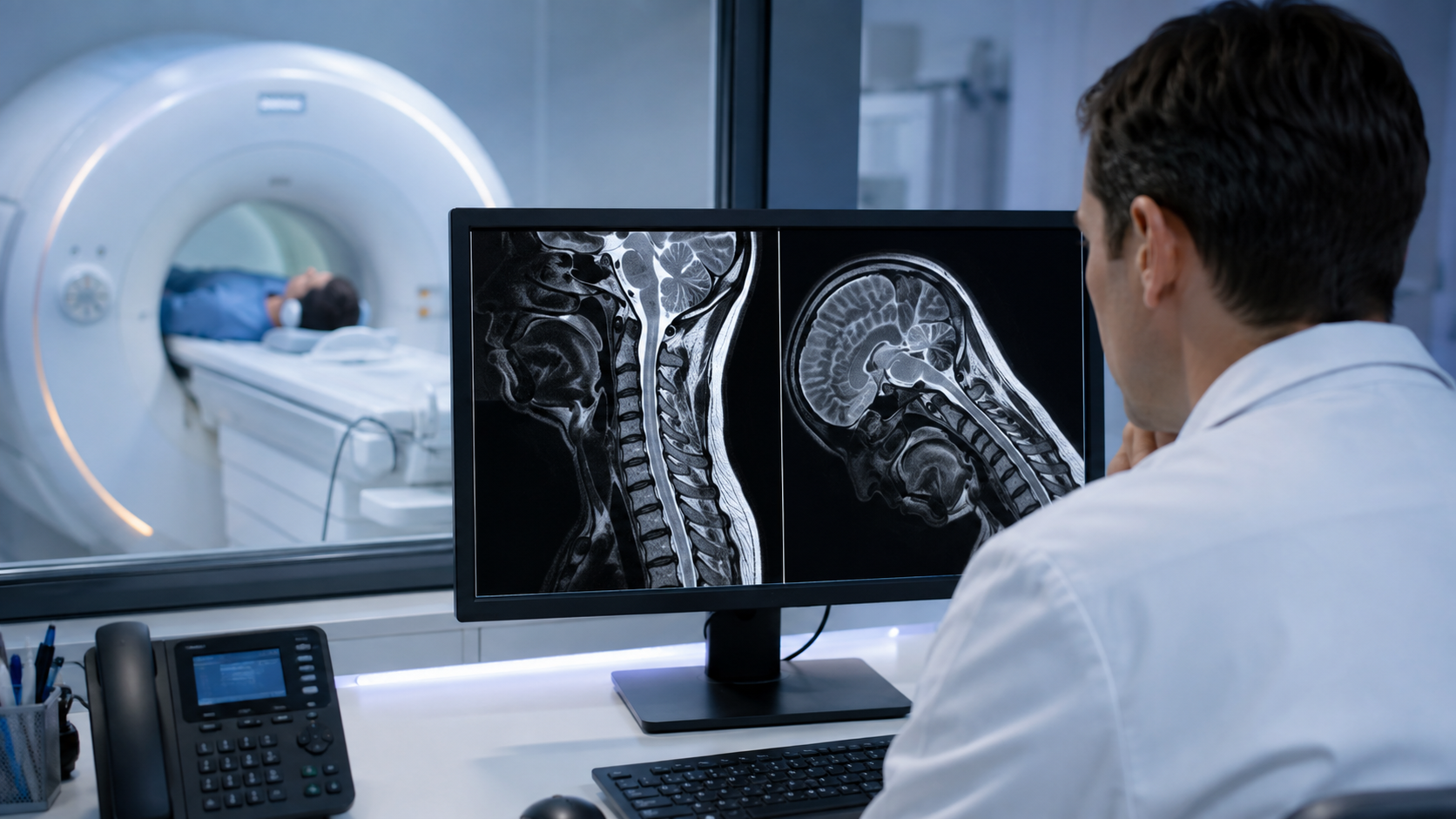
MRI, or Magnetic Resonance Imaging, uses strong magnets and radio waves to generate highly detailed images of the internal structures of your body. What makes MRI particularly useful for diagnosing arm and hand problems is its ability to capture detailed images of soft tissues—such as muscles, tendons, ligaments, and nerves—that can be difficult to see on X-rays.
Unlike X-rays, which mainly show bones, MRI can reveal issues with soft tissues, blood vessels, and even nerves. This level of detail makes it ideal for pinpointing the source of pain or dysfunction, especially when other imaging tests haven’t provided enough information.
Another benefit? MRI is completely non-invasive and doesn’t use radiation, making it a safe option for diagnosing a wide range of conditions.
When Is MRI Needed for Arm and Hand Pain?
You might be wondering when an MRI is actually necessary. Typically, doctors recommend an MRI when you have persistent or unexplained pain that hasn’t been diagnosed with other tests like X-rays or ultrasounds.
If you’ve been experiencing pain, numbness, or tingling that doesn’t go away with standard treatments, your doctor may turn to MRI to dig deeper. This is especially true for issues involving soft tissue, like tendons and ligaments, or nerve problems, such as carpal tunnel syndrome.
MRI is also a go-to test when other imaging methods fall short. For example, if an X-ray doesn’t show any obvious bone injury, but you’re still dealing with pain or loss of function, an MRI might reveal a hidden issue like a small fracture or a damaged tendon that an X-ray couldn’t detect.
Common Arm and Hand Conditions Diagnosed by MRI
MRIs are particularly useful in diagnosing several conditions that affect the arm and hand. Here are some of the most common ones:
Tendon and Ligament Injuries
Tendonitis, or inflammation of a tendon, can cause severe pain and limit movement in your arm or hand. MRI can clearly show inflamed or torn tendons that may not be visible with other tests. Ligament injuries, such as sprains or tears, are also easily spotted with MRI, giving your doctor the information they need to recommend proper treatment.
Nerve Compression and Entrapment
Conditions like carpal tunnel syndrome, where the median nerve is compressed in the wrist, can cause pain, numbness, or tingling. An
MRI can provide a detailed look at the nerve and surrounding tissues to confirm the diagnosis. Similarly, if you’re experiencing symptoms of cubital tunnel syndrome, where the ulnar nerve is compressed at the elbow—an MRI can help pinpoint the issue.
Fractures and Bone Health
While X-rays are great for detecting obvious fractures, small or hidden fractures, known as occult fractures, might not show up. MRI can detect these smaller fractures and even reveal bone bruises or stress fractures that could be causing your pain. Additionally, bone cysts or tumors, which could be contributing to discomfort, can also be diagnosed through MRI.
Arthritis and Joint Conditions
Whether you’re dealing with osteoarthritis or rheumatoid arthritis, MRI is a great tool for seeing the extent of joint damage. It can show the wear and tear on cartilage and detect inflammation around the joints. MRI is also useful in diagnosing ganglion cysts, which are fluid-filled lumps that commonly occur in the wrist and hand and may be linked to joint or tendon problems.
The MRI Process for Arm and Hand Scans
If you’ve never had an MRI before, the process can sound intimidating—but it’s actually pretty simple. There’s usually no special preparation required, although you may need to remove any metal objects like jewelry or watches before the scan.
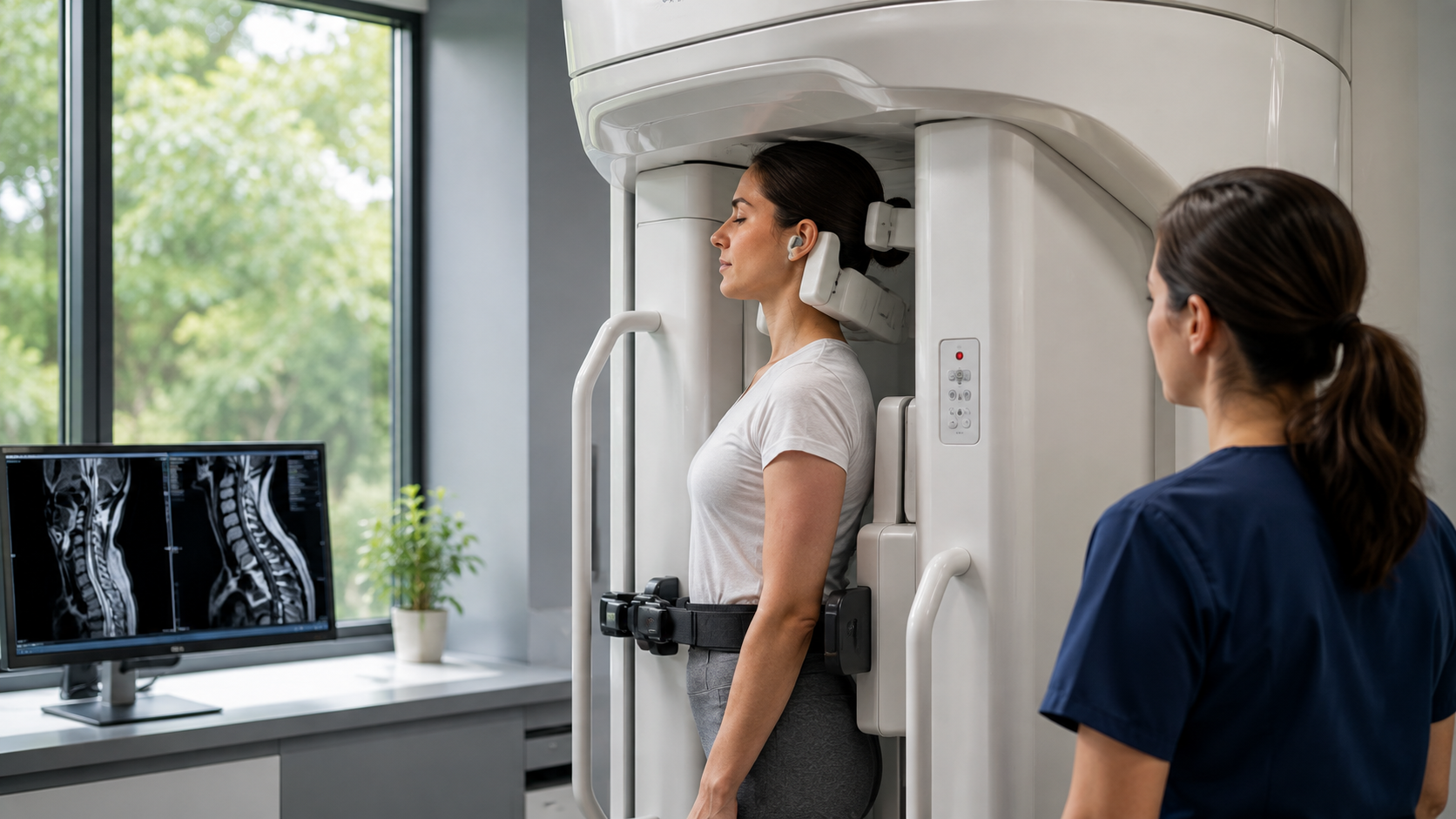
During the MRI, you’ll lie down on a table, and your arm or hand will be positioned inside the MRI machine. For most arm or hand scans, the rest of your body stays outside the machine, so it’s not as claustrophobic as you might think. The machine will make some loud noises as it takes images, but you’ll likely be given earplugs or headphones to make it more comfortable.
The scan itself usually takes 30 minutes to an hour. In some cases, contrast dye is used to make certain tissues or structures more visible, but this is usually painless and safe.
After the scan, a radiologist will review the images and send a report to your doctor, who will go over the results with you.
Understanding MRI Results and Their Role in Treatment
Once your MRI results are in, your doctor will explain what they show and how that will impact your treatment. For example, if the MRI reveals a torn tendon or ligament, your doctor might recommend physical therapy, medications, or even surgery depending on the severity of the injury.
MRI is also valuable in tracking recovery. If you’ve already had surgery or are undergoing treatment, an MRI can show how well the injury is healing and whether any complications, such as scar tissue, are developing.
Sometimes, MRI results will suggest that further testing or different treatment options are needed, giving your doctor more information to work with.
The Benefits of MRI for Arm and Hand Issues
One of the biggest benefits of MRI is how clearly it can show soft tissue injuries. This detailed imaging helps doctors avoid unnecessary procedures and provides more targeted treatment options. By catching issues early, MRI can help prevent the problem from getting worse and guide you toward the right course of treatment.
Additionally, if surgery is necessary, MRI gives surgeons a clear map of the injury so they can plan the procedure more effectively. After surgery, MRI can also be used to monitor your recovery and ensure that everything is healing properly.
What Are the Alternatives to MRI?
While MRI is a great tool for diagnosing many conditions, it’s not always the first step. X-rays are often used to rule out fractures, and ultrasound can be helpful for certain soft tissue injuries. However, these tests don’t offer the same level of detail as MRI, especially when it comes to nerve or soft tissue issues.
CT scans can be used to visualize bones, but like X-rays, they’re less effective at capturing soft tissues. MRI remains the gold standard for diagnosing complex arm and hand conditions.
Conclusion
MRI is a game-changing tool for diagnosing arm and hand issues, providing doctors with the detailed images needed to make informed treatment decisions. Whether you’re dealing with a tendon tear, nerve compression, or arthritis, MRI can illuminate what’s going on beneath the surface and guide you toward the best treatment plan.
At Upright MRI of Deerfield, we specialize in providing comfortable, high-quality MRI services to help you get the answers you need. If you’re experiencing arm or hand pain and are considering an MRI, we’re here to guide you every step of the way.
SHARE THIS POST:
Leave a Comment:

The World's Most Patient-Friendly MRI. A comfortable, stress-free, and completely reliable MRI scan. We offer patients an open, upright, standup MRI experience that helps those who are claustrophobic and stress being in a confined area. Upright MRI of Deerfield is recognized as the world leader in open MRI innovation,
Our Recent Post


READ PATIENT TESTIMONIALS
Upright MRI of Deerfield.
Susan D.,
Highland Park, 39
I am going to tell everyone about your office! This was a great experience after I panicked in other MRI machines and had to leave. Thank you so much.

Judith B.,
Milwaukee, 61
I suffer from vertigo and other MRIs do not work. This was wonderful…absolutely NO discomfort at all. The MRI was so fast…I wanted to stay and watch the movie! Mumtaz was great. His humor really put me at ease. I’ve already recommended Upright MRI to friends.
Delores P.,
Glencoe, 55
Everything is so nice and professional with your place. I have been there a couple of times. My husband and I would not go anywhere else.