Dr. Michael Fox • Nov 08, 2021

What is a Stroke and How Can MRIs Help With Detection and Recovery?


Strokes can happen unexpectedly and have the potential to leave permanent brain damage. Getting regular brain magnetic resonance imaging (MRI scans) following a stroke can help physicians track the progress of your recovery.

What is a stroke?

A stroke occurs when there is a blood flow interruption from a blood vessel to the brain, causing the brain cells to die and may cause permanent brain damage or death.


Strokes are the third leading cause of death in the United States, and most happen unexpectedly. If you or anyone you know experience symptoms, it’s critical to get medical care as soon as possible.


There are 2 types: 


Ischemic stroke: This is the most common type, and it occurs when a blood vessel is blocked from the brain due to narrowed arteries or a blood clot. 


Hemorrhagic stroke: This type occurs when a blood vessel in the brain bursts and leaks, causing bleeding into the brain. 

Symptoms of a stroke

Strokes most commonly occur unexpectedly and suddenly, but there are definite warning signs associated with someone about to experience one. If you or someone you know experiences the below symptoms, it’s critical to get medical help immediately.


  • Numbness or weakness in the face, arms or legs
  • Trouble speaking
  • Trouble seeing 
  • Dizziness
  • Trouble standing, loss of balance 

The role of MRI in strokes

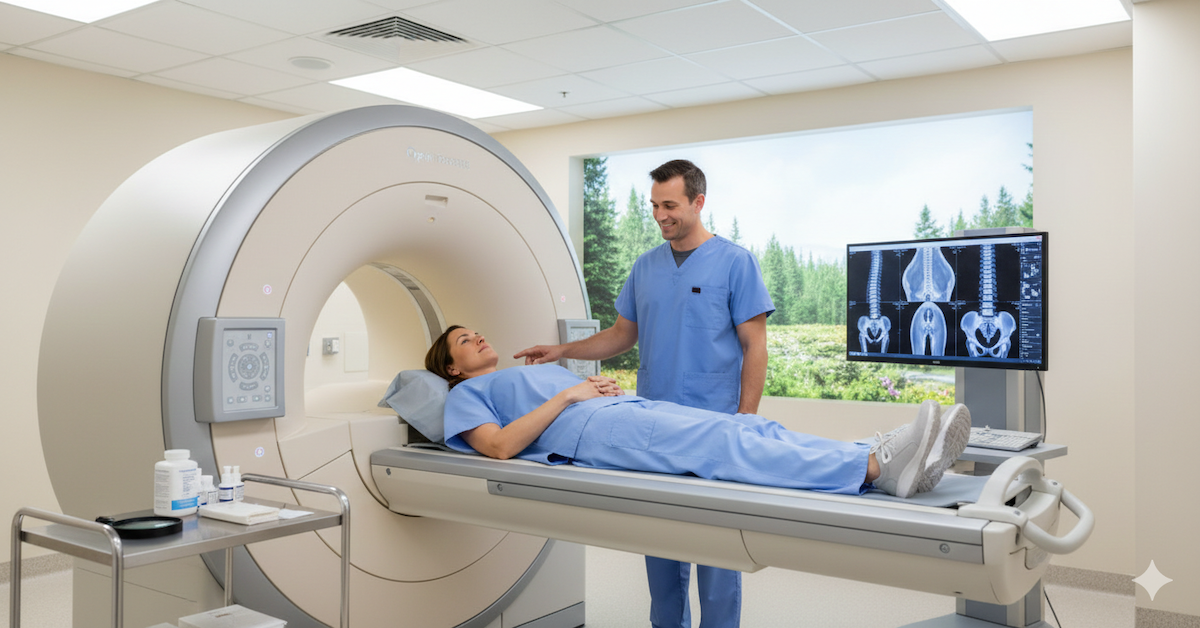
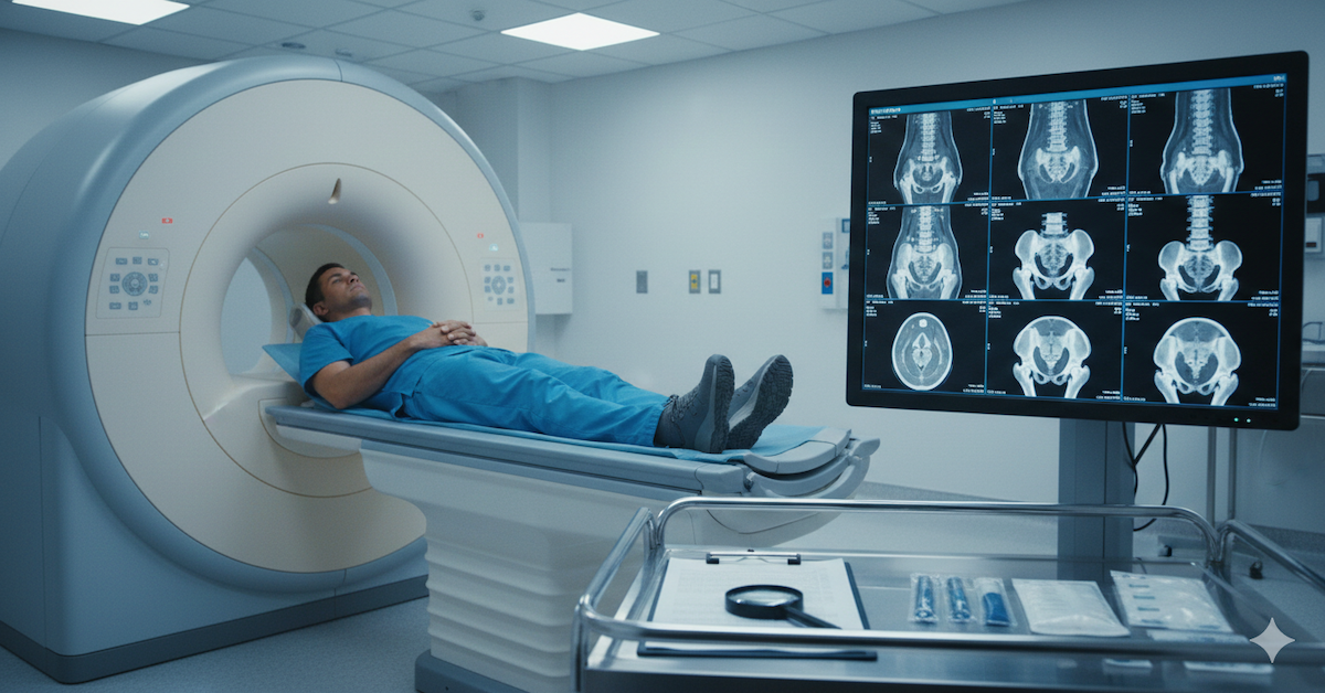
MRIs differ from other imaging scans in that they can detect specific differences between tissues that are less clear with an x-ray or CT scans.


More specifically, at Upright MRI of Deerfield, we use the Open Upright Multi-Position MRI, which is not only considered the world’s most patient-friendly MRI scanner, but also can produce very detailed images because of its ability to position patients in any position – sitting, standing, laying down or bending.


The first step following a stroke is figuring out what type it is, which is determined by getting a brain MRI and examining the damaged brain tissue. After analyzing the tissue and discussing the patient’s treatment and recovery process, the MRI is a valuable tool to track the patient’s recovery improvement over time.

How MRI can monitor recovery from stroke

The traditional process for monitoring recovery over time is with an fMRI (functional MRI) which measures the changes in blood flow that occur with brain activity. It’s also used to figure out how different parts of the brain are handling speech and mobility.


As technology advances, the MRI has proved itself to be an essential tool in stroke research and recovery, especially now with innovative scanners such as the Open Upright Multi-Position MRI machine.


To learn more about Upright MRI of Deerfield, visit our website.

SHARE THIS POST:

Leave a Comment:

Upright MRI of Deerfield

The World's Most Patient-Friendly MRI. A comfortable, stress-free, and completely reliable MRI scan. We offer patients an open, upright, standup MRI experience that helps those who are claustrophobic and stress being in a confined area. Upright MRI of Deerfield is recognized as the world leader in open MRI innovation,

LEARN MORE

Our Recent Post

MRI Brain Imaging
By Alan R 14 Apr, 2024
Prepare for peace of mind: Learn what to expect during MRI brain imaging, from pre-scan preparations to interpreting results, ensuring a smooth and informed experience.
Diagnosing Brain Conditions
By Alan R 07 Apr, 2024
Unlocking the mysteries of brain health: Discover how MRI aids in diagnosing brain conditions with precision and clarity.
What Insights Can MRI Foot Scans Provide?
By Alan R 18 Mar, 2024
Unlock valuable insights into foot health with MRI foot scans. Discover the intricate details of foot anatomy and function, aiding in the diagnosis of various conditions. I
By Alan R 06 Mar, 2024
MRI brain scans are an essential tool in modern medicine, offering detailed insights into the inner workings of the human brain. These scans serve a dual purpose: they allow healthcare professionals to visualize the anatomy of the brain with remarkable clarity, while also providing valuable information about brain function and potential abnormalities. In this article, we will explore the purpose and procedure of MRI brain scans, shedding light on why they are conducted and what patients can expect during the process. Understanding the Purpose of MRI Brain Scans At the heart of every MRI brain scan lies a simple yet profound goal: to help doctors see inside the brain without the need for invasive procedures. By harnessing the power of magnetic fields and radio waves, MRI technology generates highly detailed images of the brain's internal structures. These images can reveal important information about the size, shape, and condition of various brain regions, allowing doctors to diagnose a wide range of neurological conditions. One of the primary purposes of MRI brain scans is to detect abnormalities such as tumors, lesions, or areas of abnormal tissue growth. Unlike traditional imaging techniques like X-rays or CT scans, which provide only a limited view of the brain, MRI offers unparalleled clarity and precision. This makes it an invaluable tool for identifying potentially life-threatening conditions and guiding appropriate treatment strategies. But MRI brain scans aren't just about spotting problems they're also about understanding how the brain functions. By measuring factors like blood flow, oxygen levels, and neural activity, MRI can provide insights into brain health and performance. This information is particularly useful in cases of neurological disorders such as stroke, Alzheimer's disease, or epilepsy, where changes in brain function may precede visible structural changes.
Standard MRI
By Alan R 18 Feb, 2024
Discover the conditions overlooked by standard MRI scans. Explore the revolutionary technology that detects spinal, joint, and neurological issues in a natural, weight-bearing state.
By Alan R 11 Feb, 2024
In the realm of medical diagnostics, MRI scans have become indispensable tools for peering into the intricate landscapes of the human body. As technology advances, the traditional supine MRI, where patients lie flat, is being challenged by an innovative alternative – the Upright MRI. This article undertakes a comprehensive exploration, delving into the key distinctions between Upright and Traditional MRI scans and shedding light on how these differences can impact patient experience and diagnostic capabilities. Understanding Traditional MRI Traditional MRI, the stalwart of medical imaging, places patients in a horizontal position within a closed-bore scanner. While it has proven its worth in diagnosing various conditions, the closed design can be daunting for individuals with claustrophobia. The experience of being enclosed within the machine may result in discomfort and anxiety, potentially affecting the quality of the scan due to patient movement.
By Alan R 19 Jan, 2024
The intricate design of the human spine, while remarkable, is not exempt from the wear and tear of daily life. Spinal issues, ranging from herniated discs to spinal stenosis, can significantly impact one's mobility and overall well-being. In the realm of diagnostics, advanced tools play a pivotal role in unraveling the complexities of spinal conditions. This article takes a deep dive into the realm of Magnetic Resonance Imaging (MRI) scans, shedding light on their significance in diagnosing spinal issues. Understanding Spinal Issues Navigating life with chronic back pain or debilitating spinal conditions can be an uphill battle. From herniated discs causing discomfort to spinal stenosis affecting everyday movements , the impact is profound. Accurate and timely diagnosis becomes paramount for effective treatment. Spinal health, often taken for granted, demands attention when issues arise. Here, we explore the essential role of MRI scans in unraveling the mysteries of spinal conditions.
By Alan R 10 Jan, 2024
In the intricate landscape of medical diagnostics, Magnetic Resonance Imaging (MRI) stands out as a powerful tool, particularly when it comes to examining the complexities of the back and spine. This comprehensive guide aims to demystify the MRI process, providing patients with a deeper understanding of its significance in diagnosing various spinal conditions. If you've been recommended an MRI for your back or spine, this guide is your roadmap to navigating the procedure and interpreting the results. Understanding Back and Spine MRI Let's start by unraveling the mystery behind MRI technology. Unlike traditional X-rays or CT scans, which use ionizing radiation, MRI relies on powerful magnets and radio waves to create detailed images of the internal structures of the body. This non-invasive approach is especially beneficial for imaging the soft tissues of the back and spine , offering unparalleled clarity without exposing patients to harmful radiation.
The Importance of MRI in Diagnosing Brain Conditions
By Alan R 15 Dec, 2023
Explore the depths of diagnostic precision with our guide, highlighting the crucial role MRI plays in unveiling and understanding various neurological challenges.
Understanding MRI Foot Scans
By Alan R 02 Dec, 2023
Uncover the intricate details of foot health with our guide, highlighting the importance of MRI in understanding and diagnosing various conditions.
SEE MORE POSTS

READ PATIENT TESTIMONIALS

Upright MRI of Deerfield.


Susan D.,

Highland Park, 39

I am going to tell everyone about your office! This was a great experience after I panicked in other MRI machines and had to leave. Thank you so much.

Judith B.,

Milwaukee, 61

I suffer from vertigo and other MRIs do not work. This was wonderful…absolutely NO discomfort at all. The MRI was so fast…I wanted to stay and watch the movie! Mumtaz was great. His humor really put me at ease. I’ve already recommended Upright MRI to friends.

Delores P.,

Glencoe, 55

Everything is so nice and professional with your place. I have been there a couple of times. My husband and I would not go anywhere else.

SEE ALL TESTIMONIALS
Share by: