Dr. Michael Fox • October 25, 2021

Upright MRI Role in Detecting Tumors


What is a tumor?

A tumor is any swelling in a part of the body as a result from inflammation or a mass of tissue that builds up.


There are different types (benign and malignant) that vary on how serious they are. Benign means it is non-cancerous. Malignant means it is cancerous.

Cancerous versus benign meaning

Benign tumors have the ability to grow large, but can’t spread to other parts of the body. Malignant, or cancerous tumors, can invade nearby tissues and spread to other parts of the body through the blood and lymph systems. It’s imperative that when you are diagnosed with a malignant tumor you take action immediately to treat it.

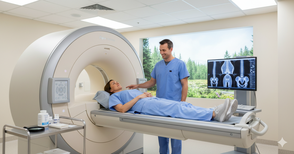
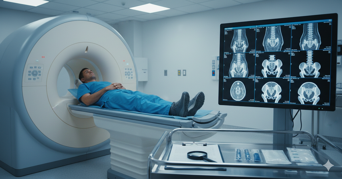
What do MRIs check for?

Magnetic resonance imaging (MRI) uses radio waves and a powerful magnet to produce detailed images of areas inside your body. This scan can create images that allows the physician to see the following:


  • If the tumor is cancerous vs. benign: The MRI is excellent at pinpointing some cancers, and can sometimes tell if a tumor is cancerous or not. 
  • The size: An MRI also allows physicians to measure the tumor, and see if it has spread. 
  • Where in the body it is: Even if the tumor is not obvious and deep inside the body, the MRI can spot its location. 
  • What a comprehensive treatment plan could be: Once the patient is diagnosed with a tumor, the detailed images that the MRI produces allows them to navigate the next best steps in treatment. 
  •  Track the effectiveness of the treatment: As the patient is treated, physicians can track their progress over time with continuous MR imaging. 


While the traditional MRI is particularly great at identifying tumors in the brain, spinal cord, and pelvic organs, the Upright MRI specifically can easily identifiable in all parts of the body due to its ability to place patients in multiple different positions.

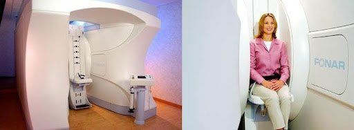
The Upright MRI advantage

The Upright MRI of Deerfield uses the FONAR Upright® Multi-Position™ MRI, which is the considered the most patient-friendly MRI in the world. While patients appreciate it for its true openness and non-claustrophobic feeling that you can’t find with traditional MRIs, it also allows physicians to position their patients in any position: Sitting, standing, laying down, or bending.


Because of these qualities, this MRI can image in greater detail, giving physicians a more accurate view of what’s happening inside the body so they can create the best treatment plan possible.


To learn more about Upright MRI of Deerfield, visit our website.

SHARE THIS POST:

Leave a Comment:

Upright MRI of Deerfield

The World's Most Patient-Friendly MRI. A comfortable, stress-free, and completely reliable MRI scan. We offer patients an open, upright, standup MRI experience that helps those who are claustrophobic and stress being in a confined area. Upright MRI of Deerfield is recognized as the world leader in open MRI innovation,

LEARN MORE

Our Recent Post

Why Is an Upright MRI Essential for Diagnosing Chiari Malformation?
By Alan R April 20, 2025
Chiari malformation is a neurological condition that can lead to serious symptoms like headaches, neck pain, and dizziness. However, diagnosing Chiari malformation is not always straightforward, especially since its symptoms can be easily mistaken for other conditions. Traditional imaging methods, such as X-rays or MRIs, sometimes fail to fully capture the complexities of the malformation. This is where Upright MRI technology comes into play. It has become an essential tool for diagnosing Chiari malformation, offering clear advantages over traditional MRI techniques by providing dynamic, functional images that help doctors better understand the condition’s impact.
Detection of Whiplash-Related Injuries
By Alan R April 13, 2025
Discover how an Upright MRI can improve the detection of whiplash-related injuries by capturing spinal alignment and soft tissue issues in weight-bearing positions.
Diagnose Cranio-Cervical Instability
By Alan R March 10, 2025
Discover how a positional MRI detects cranio-cervical instability by capturing images in different positions, revealing issues missed by traditional MRIs.
Why Are Traditional MRIs Missing Crucial Pathologies in the Spine?
By Alan R March 3, 2025
Learn why traditional MRIs may miss critical spinal pathologies and how advanced imaging techniques provide a clearer diagnosis for better treatment outcomes.
What Makes Upright MRI the Best Choice for Flexion and Extension Imaging?
By Alan R February 17, 2025
If you’ve been dealing with back or neck pain that seems to change depending on how you move, a regular MRI may not give you the answers you need. Traditional MRIs capture images while you’re lying flat, which doesn’t reflect how your spine behaves when you're sitting, standing, or bending. This is where flexion and extension imaging comes in. It allows doctors to see how your spine reacts to different movements , helping to detect issues that might not be visible in a standard scan. Upright MRI is the best option for this type of imaging because it captures the spine in a natural, weight-bearing position. Unlike traditional MRIs, which only provide a still image, an upright MRI shows how the spine changes when bending forward or backward. This can reveal hidden spinal conditions that only appear when the body is in motion.
How Can Positional MRI Reveal Hidden Spine and Shoulder Issues?
By Alan R February 7, 2025
Discover how positional MRI reveals hidden spine and shoulder conditions by capturing movement-based abnormalities missed by traditional MRIs.
What Should I Know About MRI Knee Scans?
By Alan R January 10, 2025
Learn about MRI knee scans, how they work, what conditions they detect, and how they help in creating accurate treatment plans for knee health.
By Alan R January 3, 2025
Discover how MRI imaging provides detailed insights into knee health, aiding in accurate diagnosis and effective treatment plans.
By Alan R December 5, 2024
When dealing with pelvic health issues, accurate diagnosis is critical. Pelvic conditions can be complex, involving a range of organs, tissues, and systems. That’s where MRI (Magnetic Resonance Imaging) scans come in. Known for their detailed imaging capabilities, MRIs offer a non-invasive way to assess pelvic health and pinpoint the root causes of various conditions. Here’s an in-depth look at how MRI scans can assist in diagnosing pelvic conditions and why they are a game-changer in medical imaging.
How Can MRI Scans Assist in Diagnosing Cranio-Cervical Instability?
By Alan R December 1, 2024
Cranio-cervical instability (CCI) is a condition that affects the junction where the skull meets the cervical spine. It’s a complex issue that can cause a range of symptoms, from chronic neck pain to neurological problems. Diagnosing CCI requires precision, as the condition often involves subtle changes in ligaments, bones, and other structures. MRI (Magnetic Resonance Imaging) has become a vital tool in identifying and understanding CCI, thanks to its ability to provide detailed images without invasive procedures. Here’s how MRI scans can assist in diagnosing cranio-cervical instability and why they’re an invaluable resource for patients and doctors alike.
SEE MORE POSTS

READ PATIENT TESTIMONIALS

Upright MRI of Deerfield.


Susan D.,

Highland Park, 39

I am going to tell everyone about your office! This was a great experience after I panicked in other MRI machines and had to leave. Thank you so much.

Judith B.,

Milwaukee, 61

I suffer from vertigo and other MRIs do not work. This was wonderful…absolutely NO discomfort at all. The MRI was so fast…I wanted to stay and watch the movie! Mumtaz was great. His humor really put me at ease. I’ve already recommended Upright MRI to friends.

Delores P.,

Glencoe, 55

Everything is so nice and professional with your place. I have been there a couple of times. My husband and I would not go anywhere else.

SEE ALL TESTIMONIALS