
How MRIs Can Detect Concussions

How magnetic resonance imaging (MRI) can detect and track repeated concussions over time
What is a concussion?
When you have a powerful blow to the head, whether from a sport, vehicle accident, or other traumatic head injury event, it can cause a concussion. Essentially, a concussion is when your brain moves back and forth inside your skull in rough, fast movements, which can damage your brain.
While most concussions are mild, some can be severely damaging and potentially life-threatening. They can also be harmful to your long-term health, especially if you get concussions consistently. Unfortunately, consistent concussions are typical in contact-focused sports like gymnastics or football, so it’s imperative athletes have the right coaching and equipment to practice the sport safely.

At what point do you need to get an MRI for a concussion?
It’s always best to seek medical guidance after any head injury. Physicians will typically recommend an imaging scan if your symptoms don’t go away after 48 hours, because the concussion could possibly be as severe as a skull fracture or internal bleeding. If you experience any of the following symptoms, it’s critical to get a magnetic imaging test (MRI) as soon as possible:
- Bloody nose or ears
- Blurry vision
- Confusion or agitation
- Consistent migraines
- Drowsiness or sleepiness
- Loss of consciousness
- One pupil is larger than the other
- Repeated vomiting
- Seizures
- Slurred speech
It’s essential to know that concussions can be much more severe than they may seem. A person who has a concussion may appear fine at first and be directed to just “walk it off”, but this is an extremely dangerous way to approach the situation. Brain injuries are typically not visible to the naked eye, and symptoms don’t always happen immediately after the incident.

How MRIs detect concussions
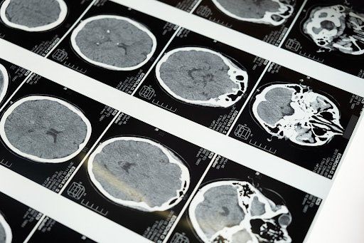
Doctors recommend patients with concussions to get brain imaging to see get an inside look at what’s happening in the brain. As technology has evolved throughout the years, MRI has become a primary imaging tool for detecting brain damages caused by concussions. They can also see if other severe issues are occurring inside the skull, like internal bleeding.
Using the body’s natural magnetic properties to create clear images of the brain tissue, MRIs can give doctors a glimpse into brain tissue damage. Nowadays, patients can receive an injection called contrast dye to show concussion-caused holes and damages to a thin membrane on the brain called meninges.
MRI and CT Scans for concussions
Patients with concussions may receive either an MRI or a CT scan to gain insight into their head injury, and some patients may need both. However, MRI can detect smaller, hard-to-see damages that a CT scan can’t, such as contusion (bruising), gliosis (scarring), and micro hemorrhage (tiny areas of bleeding).
An MRI can also be helpful when tracking the progression of the brain over time, for patients who have had many concussions. For example, many professional football players receive MRIs to track their older or repeated injuries to the brain.
Learn more about Upright MRI of Deerfield
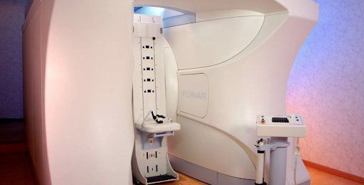
Upright MRI of Deerfield is considered the World’s Most Patient-Friendly MRI for a reason. The Upright MRI is not only completely open (patients are not enclosed and can see the front of the room); it also allows patients to be imaged in multiple positions (sitting, standing, laying down, or bending). Our MRI scan is 70% quieter than other MRIs and is specifically designed for patient comfort and its multi-position capabilities allow the scan to produce extremely detailed images that help physicians get the most accurate diagnosis possible.
To learn more about Upright MRI of Deerfield, visit our website.

SHARE THIS POST:
Leave a Comment:

The World's Most Patient-Friendly MRI. A comfortable, stress-free, and completely reliable MRI scan. We offer patients an open, upright, standup MRI experience that helps those who are claustrophobic and stress being in a confined area. Upright MRI of Deerfield is recognized as the world leader in open MRI innovation,
Our Recent Post


READ PATIENT TESTIMONIALS
Upright MRI of Deerfield.
Susan D.,
Highland Park, 39
I am going to tell everyone about your office! This was a great experience after I panicked in other MRI machines and had to leave. Thank you so much.

Judith B.,
Milwaukee, 61
I suffer from vertigo and other MRIs do not work. This was wonderful…absolutely NO discomfort at all. The MRI was so fast…I wanted to stay and watch the movie! Mumtaz was great. His humor really put me at ease. I’ve already recommended Upright MRI to friends.
Delores P.,
Glencoe, 55
Everything is so nice and professional with your place. I have been there a couple of times. My husband and I would not go anywhere else.

