
What Should I Expect from MRI Brain Imaging?
MRI brain imaging is a vital diagnostic tool used to examine the brain's internal structures and identify potential abnormalities. For many patients, the prospect of undergoing an MRI scan can be daunting. Understanding what to expect from the process can alleviate anxiety and ensure a more comfortable experience. Let's delve into the world of MRI brain imaging and explore what patients can anticipate during the procedure.
Preparing for the MRI Brain Scan
Before undergoing an MRI brain scan, there are a few important preparations to consider. Patients will typically be instructed to remove any metal objects or jewelry and to inform the healthcare provider of any medical conditions or implants. It's also essential to wear comfortable clothing without any metal components. Additionally, patients may be advised to avoid consuming food or drinks containing caffeine prior to the scan to minimize potential motion artifacts.
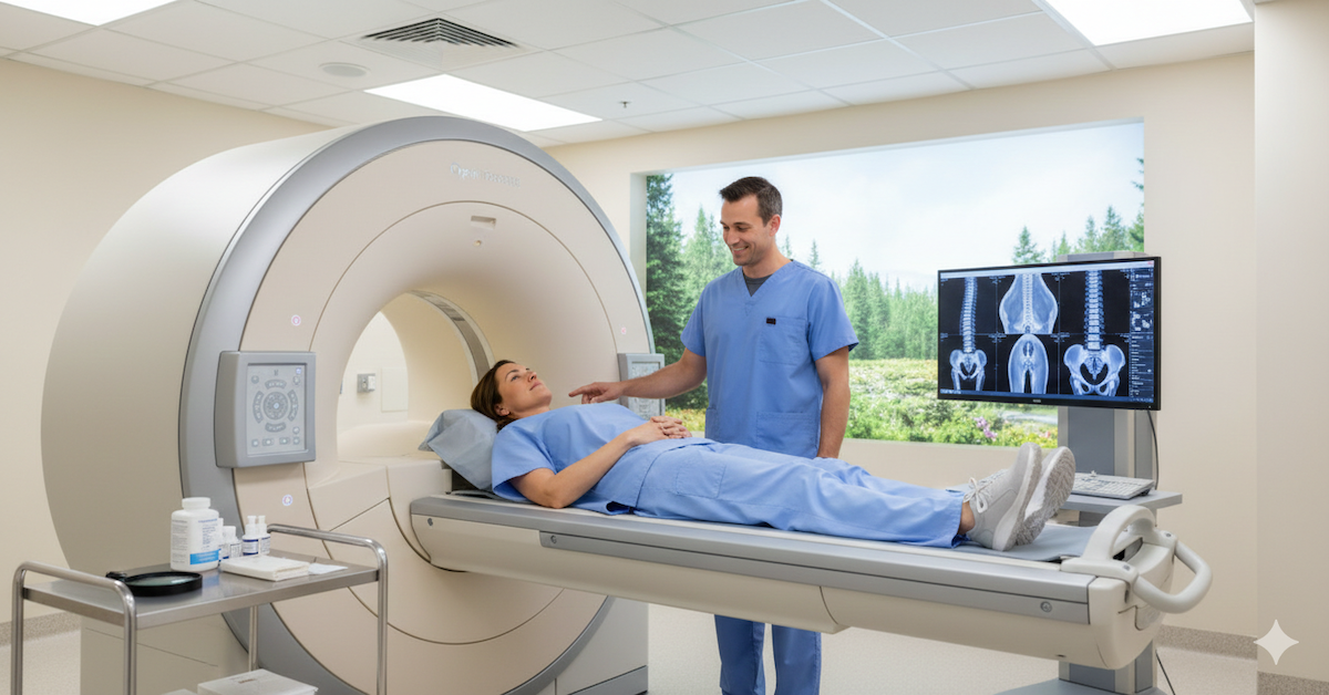
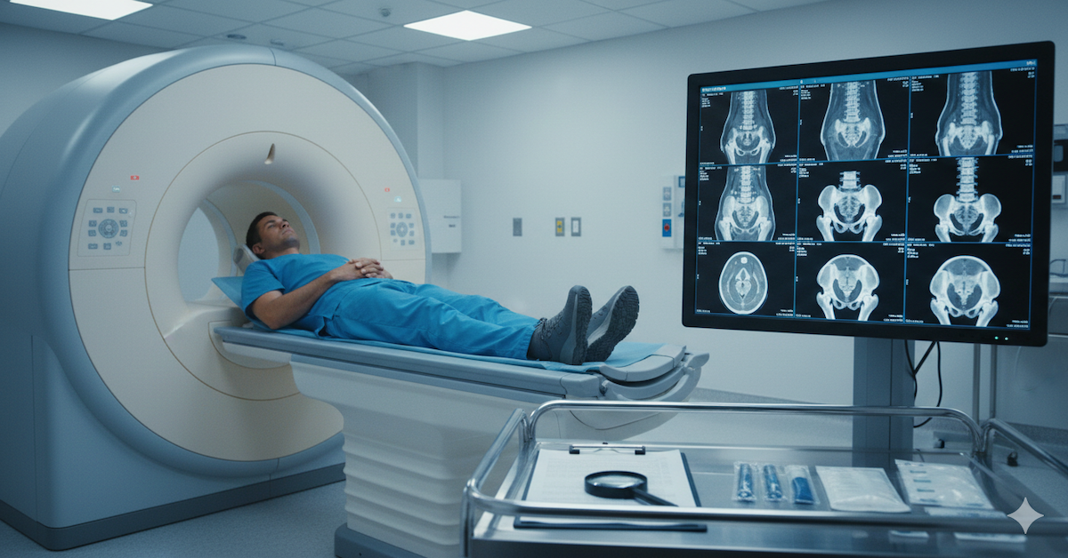
The MRI Brain Imaging Process
During the MRI brain imaging process, patients will be asked to lie still on a padded table that slides into the MRI scanner. The scanner consists of a large, tunnel-like structure surrounded by powerful magnets. While inside the scanner, patients will hear loud tapping or knocking noises as the images are being acquired. These noises are entirely normal and are a result of the magnetic field and radiofrequency pulses used in the imaging process.
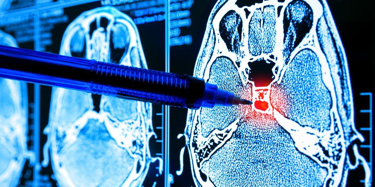
Understanding MRI Brain Images
MRI brain images are generated using different imaging sequences, each providing unique information about the brain's structures and tissues. Common types of MRI sequences include T1-weighted, T2-weighted, and FLAIR images. These images reveal detailed anatomical information, allowing healthcare providers to identify abnormalities such as tumors, strokes, or lesions within the brain. Patients may receive a copy of their MRI images and a report from the radiologist interpreting the results.
Interpreting MRI Results
Interpreting MRI results requires specialized expertise, typically provided by radiologists trained in neuroimaging. The radiologist will analyze the MRI images to identify any abnormalities or findings that may indicate a brain condition. Patients will receive a report summarizing the findings, which may include recommendations for further evaluation or treatment. It's essential to discuss the results with the referring healthcare provider to understand their implications fully.
Post-Scan Care and Follow-Up
After the MRI scan, patients can typically resume their normal activities immediately. There are no restrictions on diet or physical activity following the procedure. Patients may be advised to drink plenty of fluids to stay hydrated, especially if contrast dye was administered during the scan. It's important to follow up with the healthcare provider to discuss the MRI results and any recommended next steps, such as additional testing or treatment.
Potential Risks and Considerations
While MRI brain imaging is considered safe for most patients, there are some potential risks and considerations to be aware of. Some patients may experience claustrophobia or anxiety while inside the MRI scanner, particularly if they are prone to feeling confined in enclosed spaces. Additionally, patients with certain metallic implants or devices may not be candidates for MRI imaging. It's crucial to inform the healthcare provider of any relevant medical history or concerns beforehand.
Conclusion
In conclusion, MRI brain imaging is a valuable tool for diagnosing and evaluating a wide range of brain conditions. By understanding what to expect from the MRI process, patients can feel more informed and prepared for their scan. At Upright MRI of Deerfield, we are dedicated to providing compassionate care and accurate diagnostics to our patients. If you have any questions or concerns about MRI brain imaging, don't hesitate to reach out to our team for guidance and support.
SHARE THIS POST:
Leave a Comment:

The World's Most Patient-Friendly MRI. A comfortable, stress-free, and completely reliable MRI scan. We offer patients an open, upright, standup MRI experience that helps those who are claustrophobic and stress being in a confined area. Upright MRI of Deerfield is recognized as the world leader in open MRI innovation,
Our Recent Post


READ PATIENT TESTIMONIALS
Upright MRI of Deerfield.
Susan D.,
Highland Park, 39
I am going to tell everyone about your office! This was a great experience after I panicked in other MRI machines and had to leave. Thank you so much.

Judith B.,
Milwaukee, 61
I suffer from vertigo and other MRIs do not work. This was wonderful…absolutely NO discomfort at all. The MRI was so fast…I wanted to stay and watch the movie! Mumtaz was great. His humor really put me at ease. I’ve already recommended Upright MRI to friends.
Delores P.,
Glencoe, 55
Everything is so nice and professional with your place. I have been there a couple of times. My husband and I would not go anywhere else.

