
A Closer Look at MRI Foot Imaging: When and How it Benefits You
In the intricate dance of daily life, our feet bear the weight of our world. They navigate us through bustling streets, conquer hiking trails, and help us gracefully twirl on the dance floor. Yet, this remarkable workhorse is not immune to the wear and tear of life. From stress fractures to debilitating conditions like plantar fasciitis, foot health is paramount to maintaining an active, pain-free lifestyle. Enter MRI (Magnetic Resonance Imaging), a diagnostic marvel that delves into the intricacies of foot health, offering precise insights for diagnosis and treatment.
Understanding Foot Anatomy and Function
The foot, a masterpiece of biomechanical engineering, is a complex interplay of bones, joints, ligaments, tendons, and muscles. Bones provide structure, joints facilitate movement, and ligaments and tendons act as connectors and stabilizers. This intricate system allows us to walk, run, and perform a myriad of activities with ease. However, injuries and conditions can disrupt this delicate balance, leading to a range of foot-related issues.
MRI Technology: How It Works for Foot Imaging
MRI technology is akin to a symphony of magnets and radio waves, orchestrating a detailed performance within the body. It operates on the principle of nuclear magnetic resonance, aligning hydrogen atoms within the foot's tissues. When perturbed by radio waves, these atoms emit signals, which are then converted into high-resolution images. This non-invasive process allows for a meticulous examination of the foot's internal structures, providing a comprehensive view of any potential issues.

Advantages of MRI Foot Imaging
The advantages of MRI foot imaging are substantial. It offers a level of detail that surpasses many other imaging techniques. The high-resolution images provide a clear view of even the tiniest structures within the foot. Moreover, MRI is non-invasive, eliminating the need for surgical procedures or injections. This makes it a safer option, particularly for patients who may be averse to invasive techniques. Additionally, MRI can detect a wide range of foot injuries, from stress fractures to soft tissue issues, enabling a comprehensive assessment.
When to Consider an MRI for Foot Health
The decision to undergo an MRI for foot health is guided by symptoms and specific situations. Persistent pain, swelling, or limited mobility in the foot may warrant an MRI scan. Additionally, in cases where X-rays or other imaging methods do not provide conclusive results, an MRI becomes instrumental in obtaining a definitive diagnosis. Early diagnosis is key in preventing further complications and ensuring an optimal treatment plan.
Preparation and Procedure for an MRI of the Foot
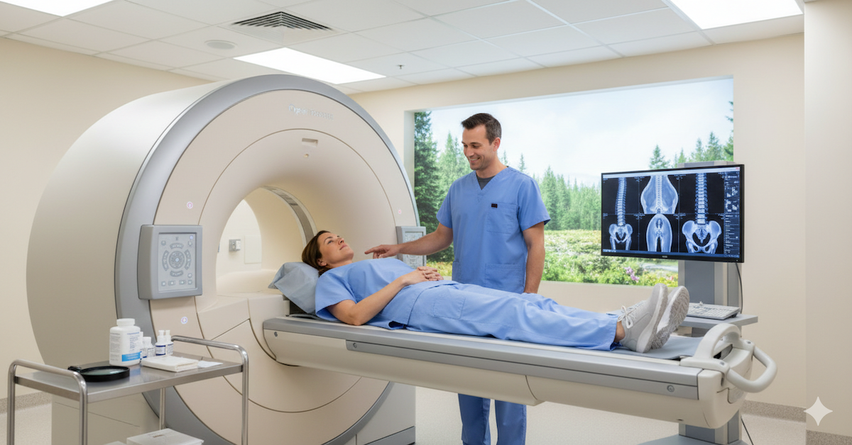
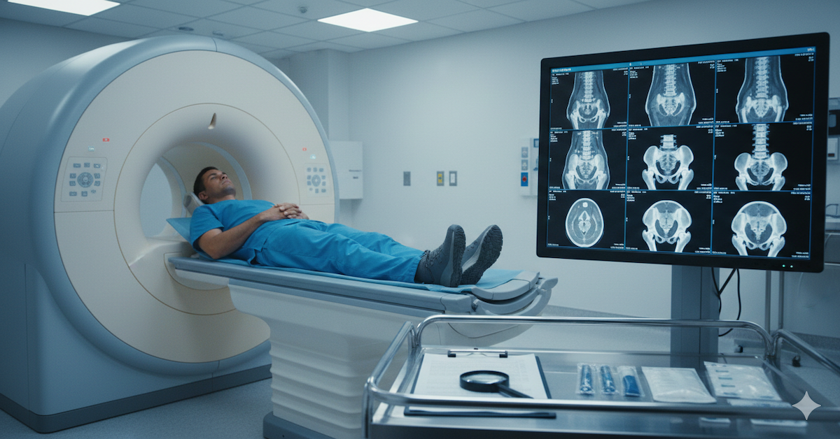
Preparing for an MRI of the foot is a straightforward process. Patients are advised to wear comfortable clothing, free of any metal. It's crucial to inform the technologist of any metal implants or devices in your body, as they can interfere with the imaging process. Once positioned comfortably, the MRI machine will emit a series of clicks and hums as it captures detailed images of your foot. The procedure is painless, but it's crucial to remain still to ensure the clarity of the images.
Common Foot Conditions Detected by MRI
MRI foot scans are incredibly versatile, capable of detecting a wide array of injuries and conditions. Stress fractures, often caused by repetitive impact, can be accurately diagnosed through MRI. Plantar fasciitis, a prevalent cause of heel pain, can be effectively assessed using this powerful imaging tool. Ligament tears, like the Lisfranc injury, can be precisely detected, guiding treatment decisions. Additionally, tendonitis and tendon tears, such as those affecting the Achilles tendon, are readily discernible through MRI scans. Even arthritis and joint disorders can be accurately evaluated, providing crucial information for treatment planning.
Interpreting MRI Results for Foot Health
Interpreting MRI results for foot health requires the expertise of a skilled radiologist with a specialization in musculoskeletal imaging. They meticulously analyze the images, identifying any anomalies or signs of injury. Patients receive a detailed report, often accompanied by visual aids like annotated images and diagrams for better comprehension. Understanding the terminology used in these reports is crucial for patients seeking clarity regarding their foot health.
How MRI Influences Treatment Plans for Foot Health
MRI doesn't stop at diagnosis; it plays a vital role in shaping treatment strategies. In cases of stress fractures, MRI guides the duration of rest required for optimal healing. For conditions like plantar fasciitis, MRI helps in tailoring physical therapy and orthotic interventions. In surgical scenarios, MRI aids in precise planning, ensuring that procedures are targeted and effective. Moreover, it serves as a compass in monitoring treatment progress, enabling adjustments based on the evolving state of the foot.
Future Trends in Foot Imaging and Treatment
As technology advances, the landscape of foot imaging and treatment is poised for remarkable progress. Emerging technologies promise even higher resolution and faster imaging, enhancing the precision of foot diagnoses. Innovations in contrast agents hold potential for greater clarity in visualizing foot structures. These developments are set to revolutionize foot health, with implications for improved patient outcomes and elevated standards of care.
Conclusion
In the intricate ballet of life, our feet are the prima ballerinas, gracefully carrying us through every step of the journey. Preserving their health and function is paramount to a vibrant, active lifestyle. MRI foot imaging stands as a beacon of hope, offering unparalleled insight into the complexities of foot conditions. With their high-resolution capabilities and non-invasive nature, MRI scans have become an indispensable tool in modern orthopedics. For individuals grappling with foot issues, seeking professional medical advice and considering an MRI scan can pave the way for a path to recovery.
At Upright MRI of Deerfield, we are dedicated to providing state-of-the-art imaging solutions to support your journey towards optimal musculoskeletal health. Visit our website here to learn more about our advanced imaging services.
SHARE THIS POST:
Leave a Comment:

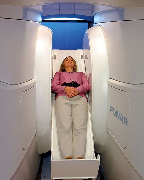
The World's Most Patient-Friendly MRI. A comfortable, stress-free, and completely reliable MRI scan. We offer patients an open, upright, standup MRI experience that helps those who are claustrophobic and stress being in a confined area. Upright MRI of Deerfield is recognized as the world leader in open MRI innovation,
Our Recent Post


READ PATIENT TESTIMONIALS
Upright MRI of Deerfield.
Susan D.,
Highland Park, 39
I am going to tell everyone about your office! This was a great experience after I panicked in other MRI machines and had to leave. Thank you so much.

Judith B.,
Milwaukee, 61
I suffer from vertigo and other MRIs do not work. This was wonderful…absolutely NO discomfort at all. The MRI was so fast…I wanted to stay and watch the movie! Mumtaz was great. His humor really put me at ease. I’ve already recommended Upright MRI to friends.
Delores P.,
Glencoe, 55
Everything is so nice and professional with your place. I have been there a couple of times. My husband and I would not go anywhere else.

