Unraveling the Diagnostic Power of MRI Back or Spine Scans: A Deep Dive
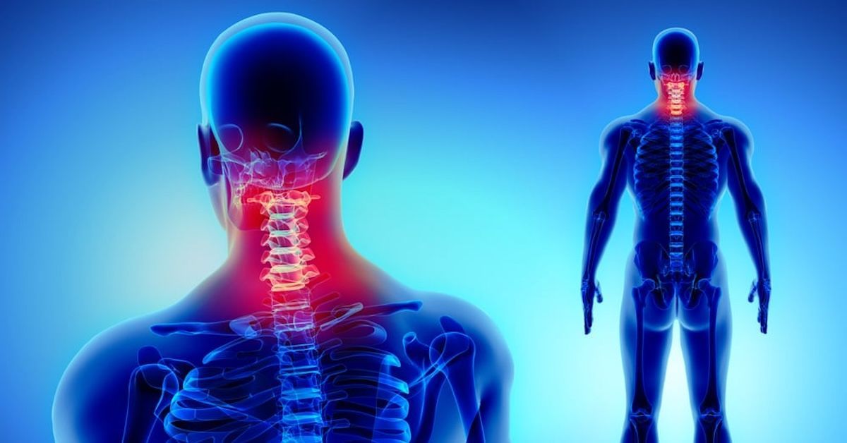
Magnetic Resonance Imaging (MRI) stands out as a potent method for identifying back and spine problems. The area of orthopedics has undergone a revolution thanks to its capability to provide precise pictures of soft tissues, bones, and nerves. We will examine the numerous illnesses it may diagnose, examine the complexities of MRI technology, and throw light on the planning and interpretation procedures in this extensive manual. Let's thus start our adventure through the complex world of MRI scans for back and spine problems.New Paragraph
Knowing about MRI technology
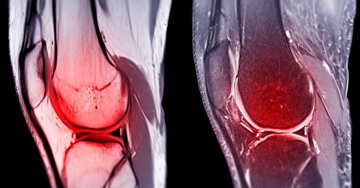
Strong magnetic fields and radio waves are used in magnetic resonance imaging, or MRI, to provide precise pictures of inside structures. MRI uses no ionizing radiation, making it a safer alternative for repeated imaging than X-rays or CT scans. This technology provides a three-dimensional view, allowing healthcare professionals to examine the spine in exquisite detail.
Types of Back and Spine Issues Diagnosed by MRI
Herniated Discs
A herniated disc occurs when the soft tissue inside the spinal disc protrudes through the tougher exterior. This can lead to nerve compression and intense pain. MRI is particularly adept at capturing these anomalies, allowing for accurate diagnosis and assessment of their severity. It aids in determining the best course of treatment, be it conservative methods or surgical intervention.
Spinal Stenosis
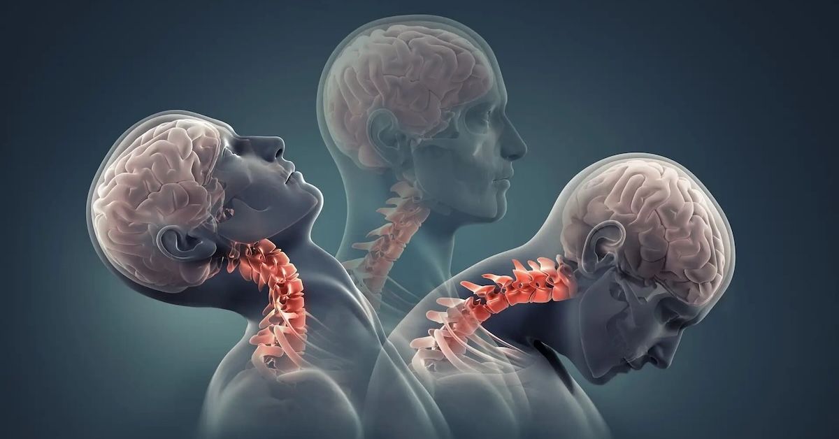
Spinal stenosis is characterized by the narrowing of the spinal canal, often leading to pressure on the spinal cord or nerves. Through MRI, the extent of this narrowing can be precisely measured. This knowledge is essential for creating treatment plans, which might include everything from surgical treatments to physical therapy to relieve pressure on the damaged tissues.
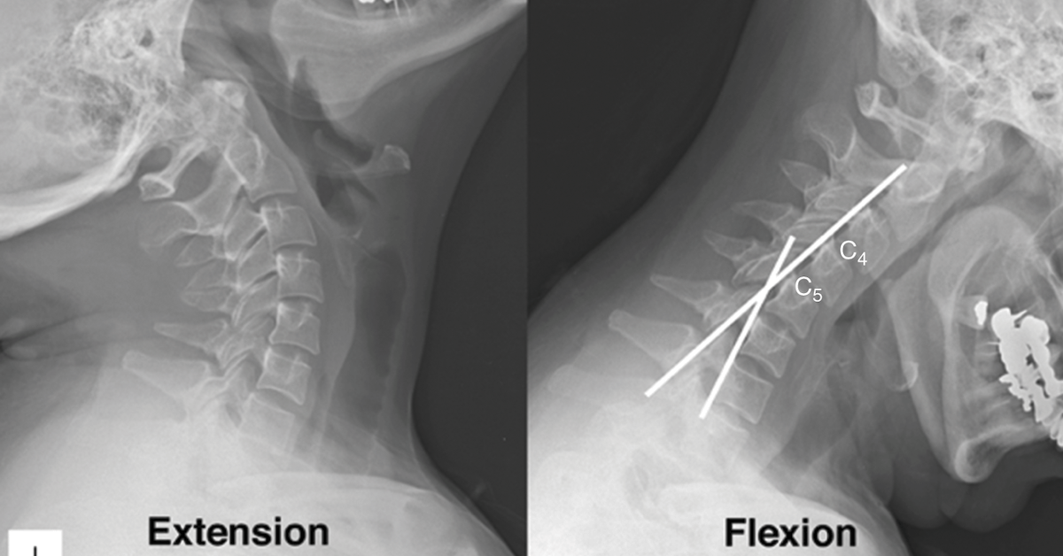
Spondylolisthesis
A condition known as spondylolisthesis occurs when a vertebra slides forward or backward in relation to the surrounding vertebrae. With the use of MRI, this displacement may be properly graded and observed. Knowing the extent of slippage is essential for selecting the best course of treatment, which may involve surgery, physical therapy, or bracing.
Degenerative Disc Disease
Degenerative disc disease can develop as we age as a result of wear and tear on the discs that support our spine. MRI is helpful in identifying these modifications and presenting a clear picture of the condition's development. With this knowledge, medical professionals may create treatment regimens that may include techniques for pain management, physical therapy, or in extreme circumstances, surgery.
Tumors and Abnormal Growths
MRI is essential for the early identification and characterisation of cancers and other aberrant spine growths. It offers thorough photos that aid in locating, sizing, and characterizing these abnormalities. Planning surgical operations and selecting the next step in treatment, such as chemotherapy or radiation therapy, both require access to this information.
Injuries and Trauma
In cases of back or spine injuries, timely and accurate diagnosis is crucial for initiating the appropriate treatment.
MRI excels in this regard, allowing healthcare professionals to assess the extent of injuries, including fractures, ligament tears, and spinal cord damage. Armed with this knowledge, they can make informed decisions regarding surgical intervention, immobilization, or rehabilitation.
Preparation for an MRI Back or Spine Scan
Patients should be informed of a few crucial procedures before having an MRI. Dietary limitations, the requirement to remove metallic items, and the significance of presenting proper medical history are a few examples. Any sensitivities or prior negative responses to contrast agents must be disclosed, if necessary. This guarantees a quick and secure imaging procedure.
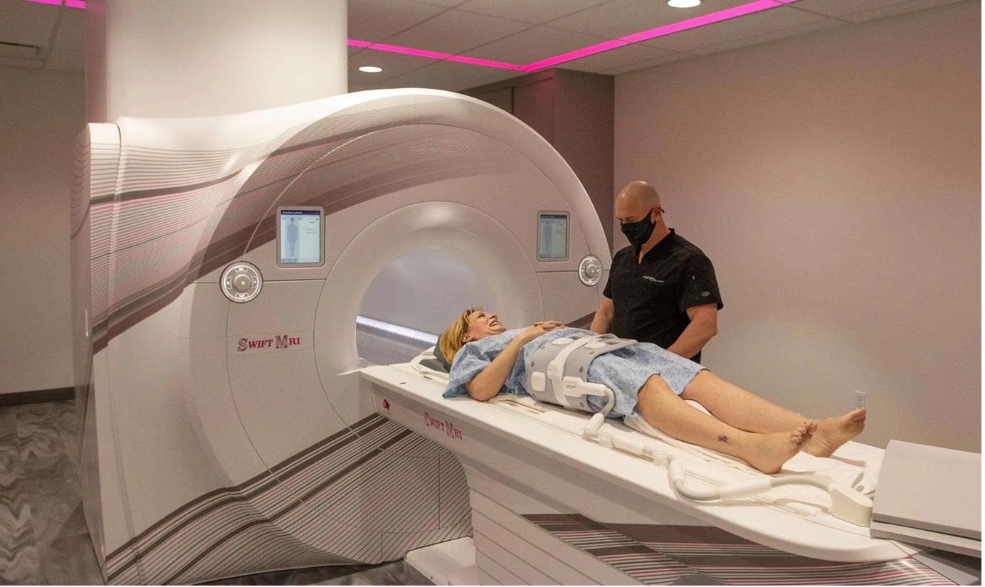
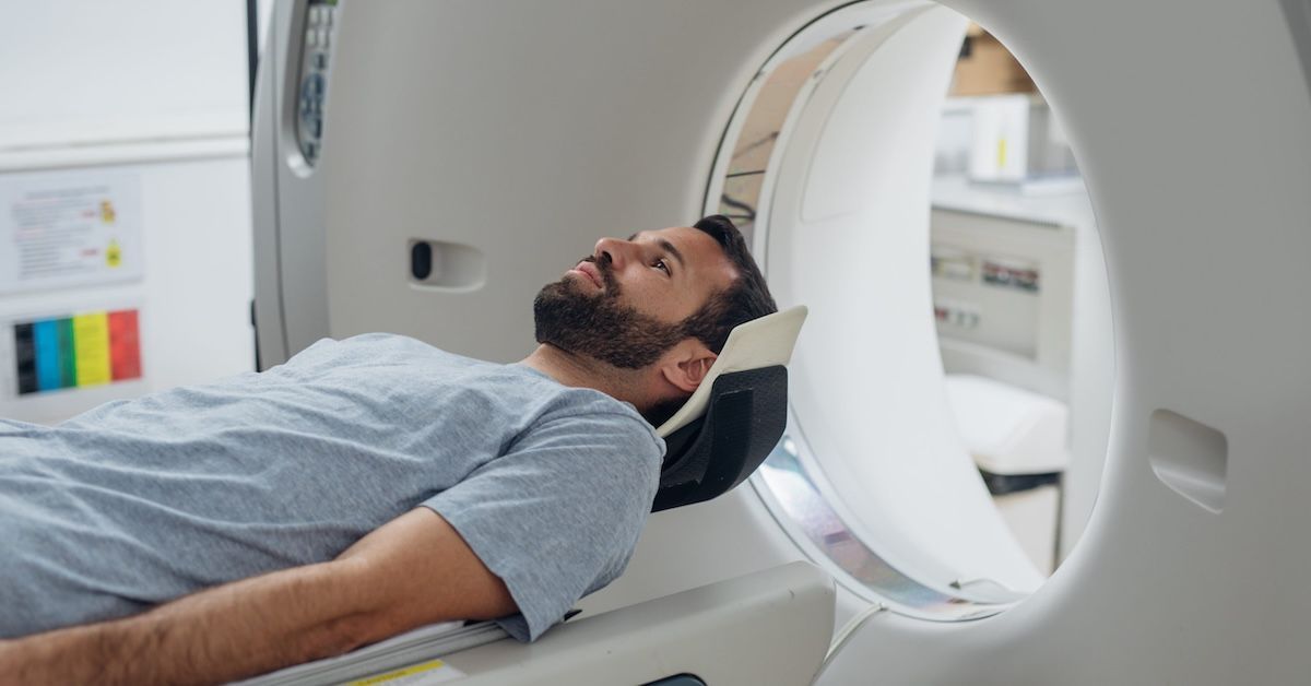
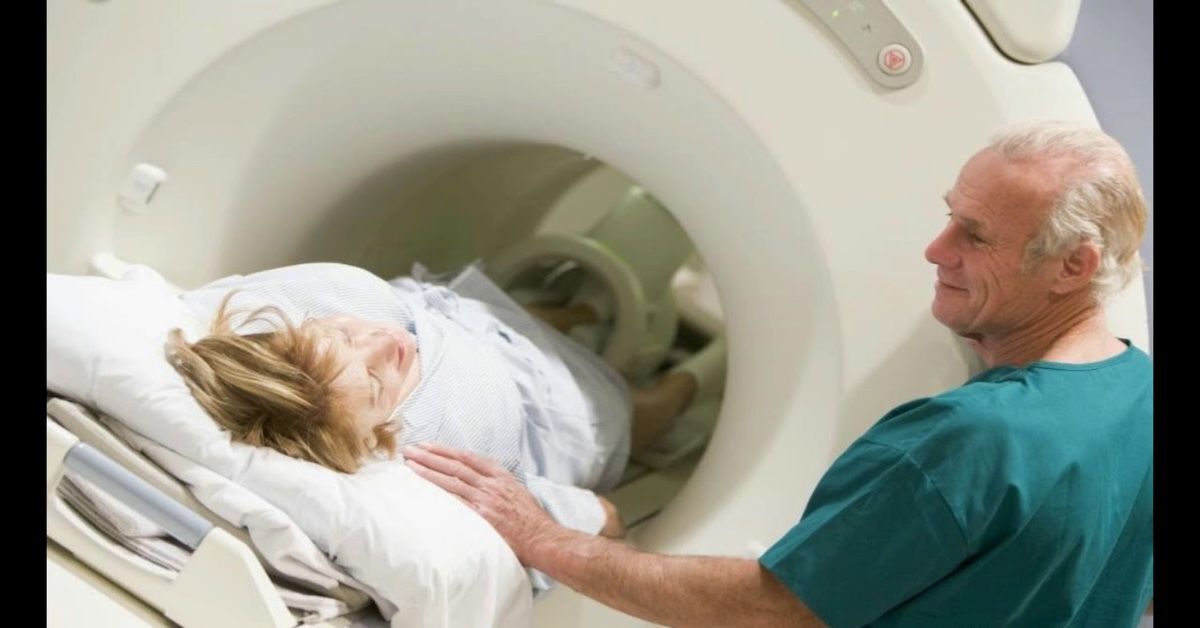
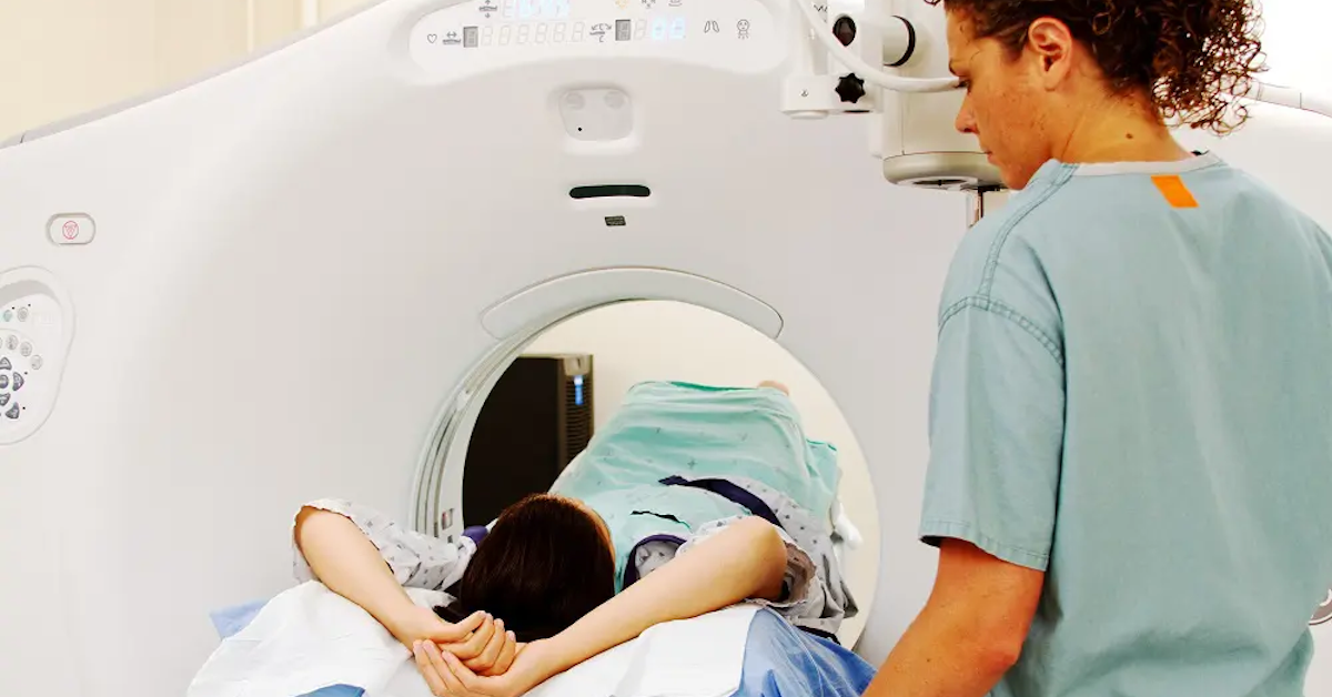
The MRI Process
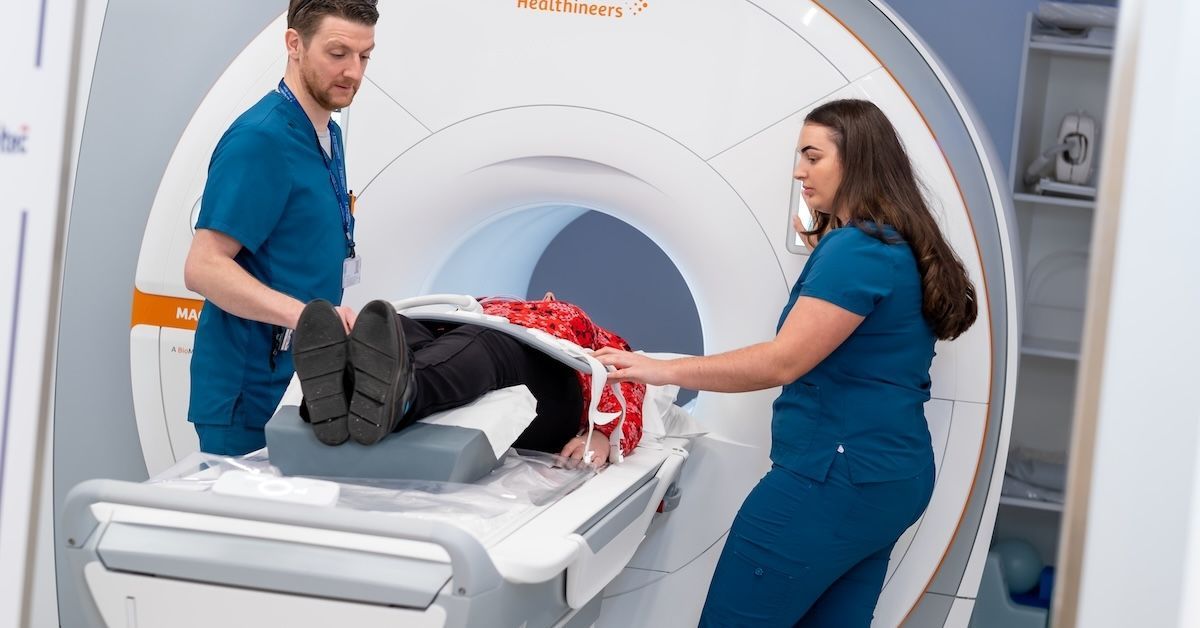
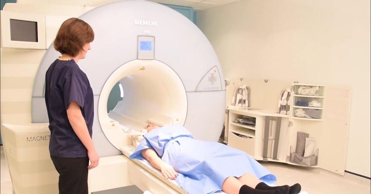
The MRI technique itself is non-invasive and painless. On a comfy table that glides inside the MRI unit, patients lie down. To achieve precise and clear pictures during the scan, it's crucial to maintain your stillness. A radiologic technologist will be in close contact with the patient the entire time, giving directions and seeing to their comfort. Depending on how detailed the examination is, the scan might take anywhere from 30 minutes to over an hour.
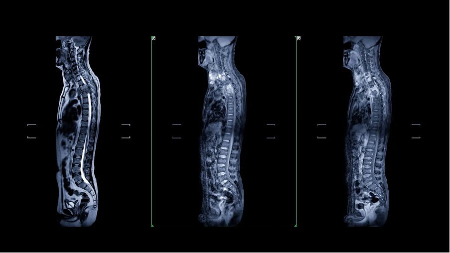
Interpreting MRI Results
After being acquired, the MRI pictures are thoroughly scrutinized by radiologists. These specialists search for certain signs of the ailment they are evaluating. The referring physician receives detailed reports that provide important information. Although some medical language may be involved in understanding these reports, your healthcare professional will walk you through the results and how they may affect your treatment strategy.

Benefits and Limitations of MRI for Back and Spine Diagnoses
There are several benefits to using MRI to diagnose problems with the spine and back. It is frequently the favored option since it can produce high-resolution pictures of soft tissues without the use of ionizing radiation. It's crucial to remember that there are instances in which different imaging modalities, such X-rays or CT scans, may be more appropriate. Based on your unique situation, your healthcare professional will decide what is the best course of action.
Future Trends in Back and Spine Imaging
Back and spine imaging is a discipline that is developing along with technology. New approaches offer scan times that are shorter and photos with even greater quality. Furthermore, advancements in imaging techniques and contrast agents have the potential to substantially improve diagnostic precision. Keeping up with these advancements can help patients and healthcare professionals make the best choices for their treatment.
Conclusion
When it comes to the diagnosis of back and spine problems, magnetic resonance imaging has shown to be revolutionary. It enables accurate and early identification of a variety of disorders thanks to its capacity to generate three-dimensional, detailed pictures of interior structures. Healthcare professionals may customize treatment strategies to meet the particular needs of each patient with prompt and precise diagnosis. To ensure the greatest outcome for your spine health, keep in mind that your healthcare team, including Upright MRI of Deerfield, is here to assist you at every stage of this process. Accept the power of information and take initiative to move toward a pain-free, healthier future.
SHARE THIS POST:
Leave a Comment:

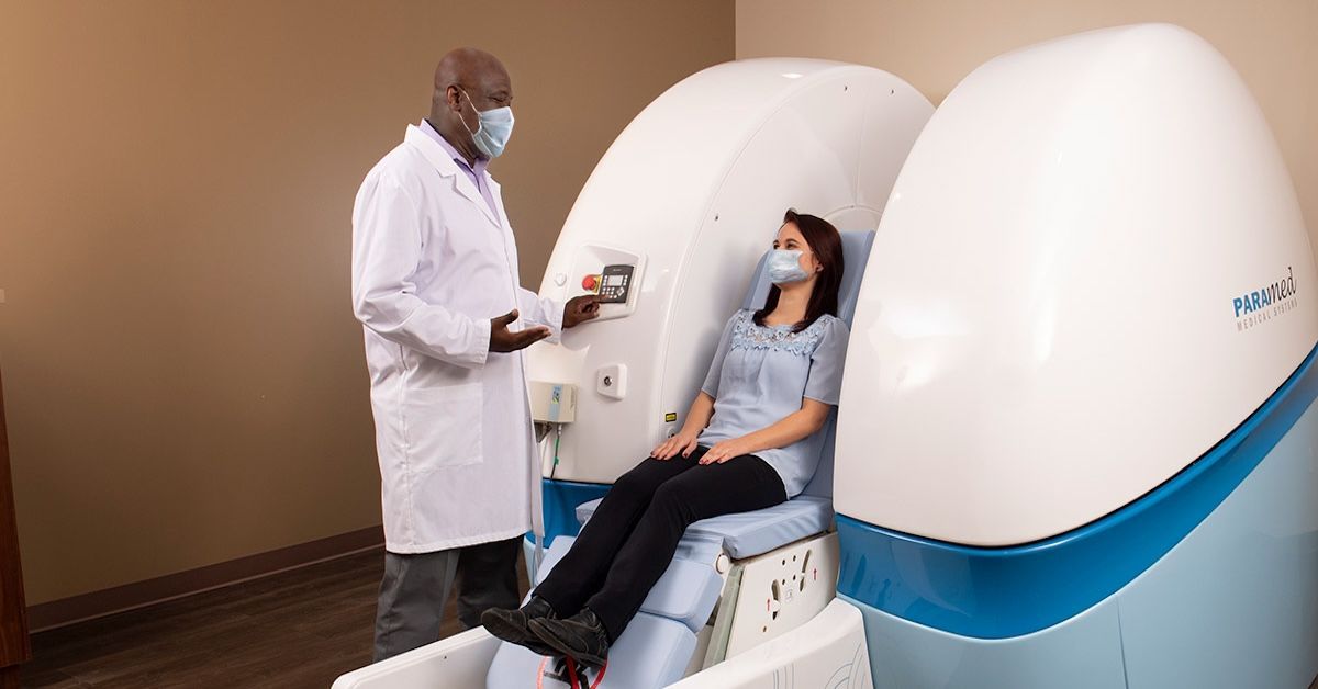
The World's Most Patient-Friendly MRI. A comfortable, stress-free, and completely reliable MRI scan. We offer patients an open, upright, standup MRI experience that helps those who are claustrophobic and stress being in a confined area. Upright MRI of Deerfield is recognized as the world leader in open MRI innovation,
Our Recent Post
READ PATIENT TESTIMONIALS
Upright MRI of Deerfield.
Susan D.,
Highland Park, 39
I am going to tell everyone about your office! This was a great experience after I panicked in other MRI machines and had to leave. Thank you so much.

Judith B.,
Milwaukee, 61
I suffer from vertigo and other MRIs do not work. This was wonderful…absolutely NO discomfort at all. The MRI was so fast…I wanted to stay and watch the movie! Mumtaz was great. His humor really put me at ease. I’ve already recommended Upright MRI to friends.
Delores P.,
Glencoe, 55
Everything is so nice and professional with your place. I have been there a couple of times. My husband and I would not go anywhere else.


